The renal system, also known as the urinary system, consists of the urethra, bladder, ureters, and the kidneys.
Overview
The kidneys are two bean-shaped organs responsible for filtering minerals from the blood, maintaining overall fluid balance, excreting waste products, and regulating blood volume to name a few.
The kidneys are very important organs in the body. About one-third of all blood leaving the heart passes into the kidneys for filtration before being pumped to the cells and tissues of the body. When the kidneys malfunction, or if they stop working (kidney failure), it may lead to various complications such as fluid retention that can lead to edema or swelling of the extremities, pulmonary edema or fluid in the lungs, hyperkalemia or increased potassium levels in the blood, anemia, heart disease, and pericarditis, among others.
Anatomy of the Kidney
Location
The kidneys are located on either side of the spine, in the retroperitoneal space. The left kidney is situated a little higher than the right one, because of the liver on the right side of the abdominal cavity, above the right kidney.
Structure
Each of the two bean-shaped organs weighs about 125 to 175 grams and 115 to 155 grams in males and females respectively. The kidney typically measures approximately 11 to 14 centimeters in length, 6 centimeters in width and is about 4 centimeters thick.
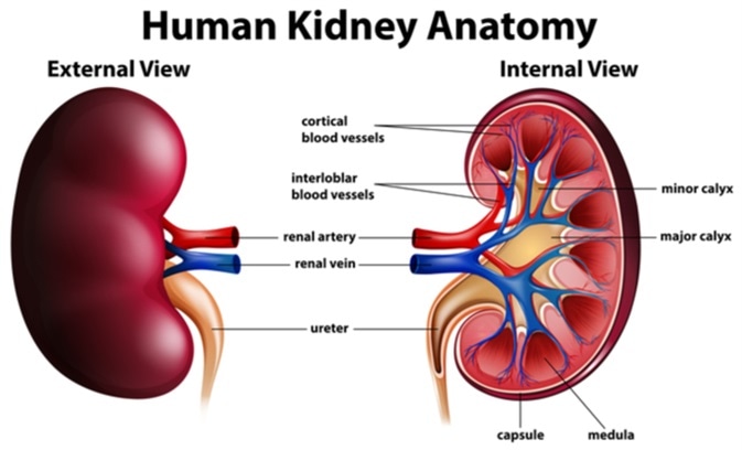
The kidneys are protected by fat, muscles, and ribs of the back. Perirenal fat, also called the renal fat pad, protects the kidneys from external force or damage. The kidneys have a medial dimple called the renal hilum, which is the entry and exit point for structures that supply or drain the kidneys such as the nerves, ureters, vessels, and lymphatics.

Vascular Supply
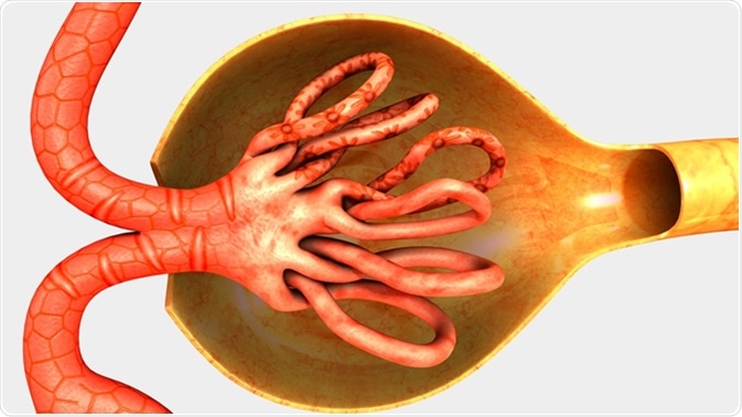
The kidneys are supplied with blood by the renal arteries, direct major branches of the abdominal aorta. These enter and divide into multiple levels to form a highly specialized and uniform network of afferent arterioles that then form the specialized capillary beds called the glomeruli. Each glomerulus forms one component of a nephron. The capillaries then merge again to form the efferent arterioles. In the outer cortex, these efferent arterioles twine around the tubules to form a peritubular network. In the inner third of the cortex and medulla, the peritubular network is replaced by long straight branches called the vasa recta.
The filtered blood travels through the left and right renal veins which empty into the inferior vena cava to reach the heart.
Parts of the Kidney
Nephrons
The nephrons are the functional units of the kidneys, and number about 1.3 million per kidney. A nephron has two main parts – tubules and corpuscles. The cup-shaped corpuscles contain the glomeruli. The tubules are small tubes traveling through the inner part of the kidney that regulate the passage of various chemicals to and from the blood. They have three parts, two highly coiled parts separated by a U-shaped curve (like a hairpin), called the proximal convoluted tubule, the loop of Henle and the distal convoluted tubule.

Other Parts of the Kidney
The cortex is the outer renal tissue located under the renal capsule. It appears lighter in color compared to the other parts of the kidney. The renal corpuscles, as well as the proximal and distal convoluted tubules are found here. It reaches into the inner renal tissue or medulla, splitting it into triangular parts called renal pyramids.
The renal pyramids contain the loops of Henle of each renal tubule, as well as the collecting ducts. Each of the renal pyramids opens at the apex, or renal papilla. Here the urine formed inside the pyramids is collected through a structure called the minor calyx. Several of the minor calices join to form a major calyx. Urine moves through the major calices to reach the renal pelvis. The pelvis is a funnel-shaped collecting structure formed by the junction of all the major calices. It carries urine from the whole kidney to the ureter, which begins where the pelvis ends, at the pelvi-ureteral junction.
The Gerota’s fascia is a thin fibrous tissue located outside the kidney and also enclosing the adrenal glands sitting atop the kidneys. It anchors the organ to the posterior abdominal wall. The renal capsule is a layer of tough fibrous tissue surrounding the kidney, found just inside the renal fat pad.
Sources
- www.niddk.nih.gov/…/kidneys-how-they-work
- https://www.niddk.nih.gov/health-information/kidney-disease
- www.betterhealth.vic.gov.au/health/conditionsandtreatments/kidneys
- https://www.ncbi.nlm.nih.gov/books/NBK459158/
Further Reading
- All Anatomy Content
- All Kidney Content
- Where are the Kidneys and Liver Located?
- Renal Neoplasms
- All Nephrology Content
Last Updated: Oct 24, 2018

Written by
Angela Betsaida B. Laguipo
Angela is a nurse by profession and a writer by heart. She graduated with honors (Cum Laude) for her Bachelor of Nursing degree at the University of Baguio, Philippines. She is currently completing her Master's Degree where she specialized in Maternal and Child Nursing and worked as a clinical instructor and educator in the School of Nursing at the University of Baguio.
Source: Read Full Article
