
The Intelligent Medical Imaging (iMED) Group at the Cixi Institute of Biomedical Engineering, Ningbo Institute of Materials Technology and Engineering (CNITECH) of the Chinese Academy of Sciences proposed a fully automated method for image-level corneal nerve fiber tortuosity estimation, contributing to the examination and diagnosis of eye-related diseases. The study was published in IEEE Transactions on Medical Imaging.
Existing clinical research suggests that the morphological changes in anatomical curvilinear structures, such as blood vessels or nerve fibers, are closely linked to various diseases. Tortuosity is one of the most significant biomarkers reflecting variations in corneal nerve fibers, serving as a significant clinical parameter to assess eye-related diseases, like hypertensive retinopathy and diabetic neuropathy.
However, there is no universally-accepted standard measure of tortuosity. Meanwhile, traditional automated tortuosity estimation relies heavily on the quality of segmentation results, which may generate errors due to poor imaging quality and low image resolution.
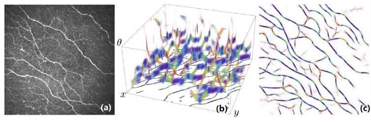
To address this issue, the research group proposed a new automated method for image-level tortuosity estimation, comprising image enhancement, exponential curvature estimation, and tortuosity level classification. They proposed an extended noise-constrained Retinex model to enhance corneal confocal microscopy (CCM) images, which was able to correct imbalanced illumination, so as to improve image contrast. By virtue of the exponential curvature estimation in the 3-D space of positions and orientations, they were able to directly measure curvature based on the enhanced images, rather than relying on the explicit segmentation and skeletonization steps in a conventional pipeline.
Source: Read Full Article