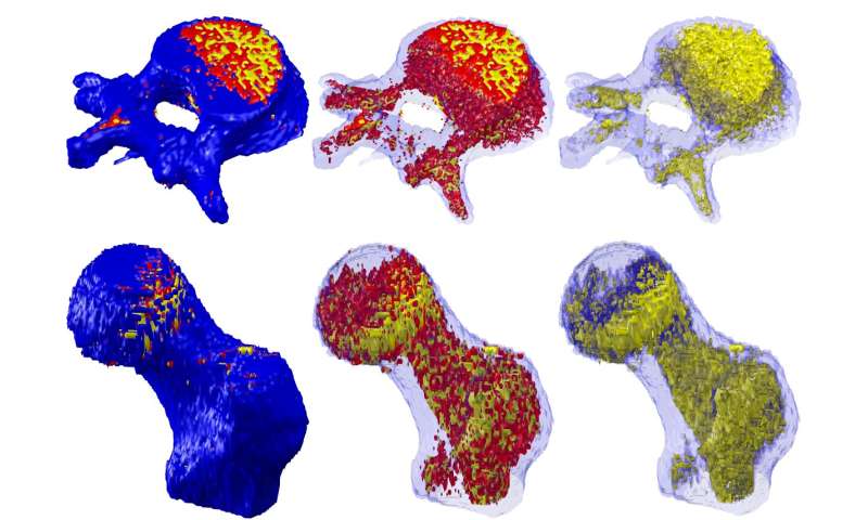
Doctors will soon be able to use fat to diagnose musculoskeletal disease within seconds and predict the risk of falls and fractures in older people, thanks to a world-leading tool developed by Melbourne researchers.
Among the diseases are sarcopenia (muscle loss), osteoporosis (brittle bones) and osteosarcopenia—a newly described syndrome encompassing osteoporosis and sarcopenia.
Two in three older Australians live with these chronic musculoskeletal conditions, which are largely associated with aging and can lead to decreased muscle mass, physical disability and poor quality of life.
Tissue Compass was developed by University of Melbourne researchers at Western Health and the Australian Institute for Musculoskeletal Science (AIMSS) to enable clinicians to quantify, within seconds, fat infiltration of muscle and bone.
“Fat infiltration in bone and muscle is the clearest indicator we have of osteosarcopenia and can be identified much earlier and accurately than changes to bone and muscle mass, which are used by doctors at the moment to make a diagnosis,” University of Melbourne Professor Gustavo Duque said.
“These local fat levels in muscle and bone herald a range of conditions that are associated with osteosarcopenia and mostly respond very well to early treatment, such as kidney disease, vitamin D deficiency, and in men, testosterone loss. They’re also associated with fractures and falls. This tool will give clinicians the information they need to diagnose osteosarcopenia and to act now to protect their patients’ health and independence into extreme old age.”
Led by Dr. Ebrahim Bani Hassan, a musculoskeletal pathophysiologist, and Mahdi Imani a biomedical engineer and masters student at the University of Melbourne, the team is now testing Tissue Compass in more than 3000 participants, in collaboration with six top American universities.
This will complete the validation of Tissue Compass in humans, which will soon become a diagnostic tool for osteosarcopenia.
Despite its prevalence, researchers say awareness of sarcopenia especially, even among doctors, is low, partly due to scant guidance on what constitutes normal versus pathological mass and function loss, and how to treat it at different stages.
Clinicians have traditionally used a combination of imaging (MRIs, CT scans and densitometries) to assess muscle and bone mass, as well as grip and gait tests. Tissue Compass can be used with routine CT and MRI scans, requiring no additional imaging.
The team unveiled the tool at last month’s World Congress of Osteoporosis, reporting that Tissue Compass is an easier, faster and more consistent tool to quantify musculoskeletal tissues and their components than usual image analysis techniques.
“There is plenty we can do through diet, exercise and medication to build up bone and muscle strength, years before people have their first fracture or fall—a moment when things can very abruptly reach a point of no return,” Professor Duque said.
Source: Read Full Article
