
Introduction
Causes and Symptoms
Subtypes of ACC
Epidemiology
Diagnosis and Treatment
References
Further Reading
Aplasia cutis congenita or ACC is a rare congenital condition. It is marked by the absence of specific layers of skin in some parts of the infant's body. It most often affects the scalp but can also affect the trunk, arms, and legs. A thin, translucent membrane is usually applied to the affected area. It's possible that the skull and/or beneath parts are visible, and that they're abnormally developed. Aplasia cutis congenita, first described by M. Cordon in 1767, manifests as a single defect on the scalp in 70% of cases, but it can also manifest as numerous lesions.
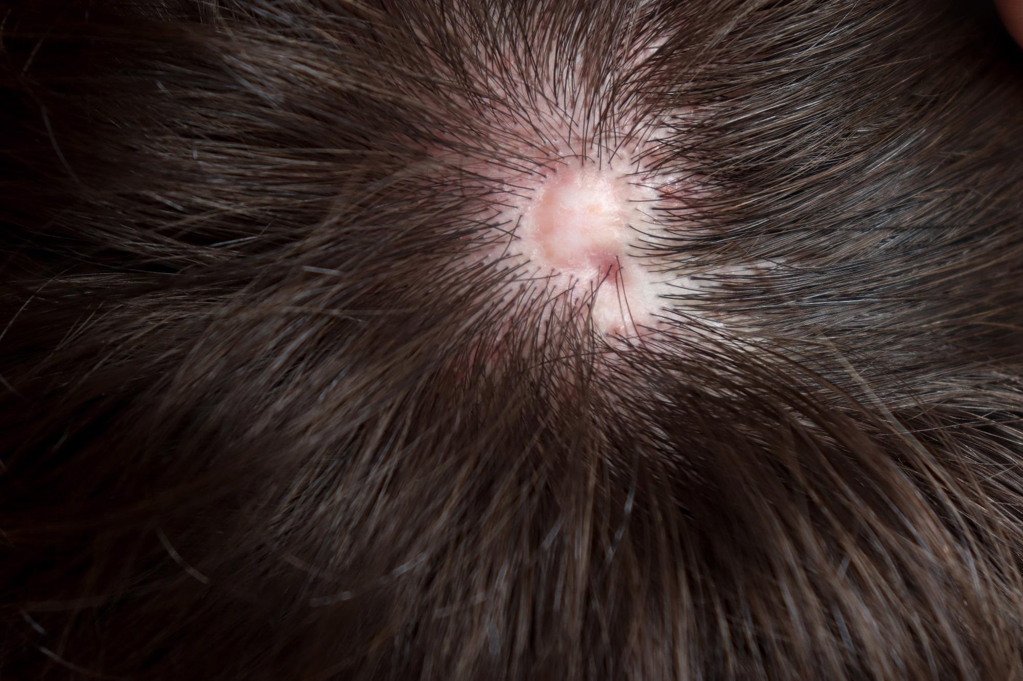 Nonsyndromic aplasia cutis congenita, a congenital defect with an absence of the epidermis, dermis and sometimes subcutaneous tissues. Usually on the top of the head. Image Credit: Kari Ahlers / Shutterstock
Causes and Symptoms
Aplasia Cutis Congenita is a rare hereditary condition that can be autosomal dominant or autosomal recessive. ACC can be caused by various factors, including familial cases, epidermolysis bullosa (EB), and imperfect neural tube closure. Ulcers can be superficial or deep at birth, obliterating all layers of skin and, in some cases, spreading to the dura or bone. Due to in utero healing, a healed scar may be the only discovery in some situations. Understanding the pathogenesis of ACC is critical in the diagnosis and therapy of these patients due to its diverse presentation.
The specific pathophysiology of ACC is yet unknown. Intrauterine trauma, vascular impairment, infectious agents, and drugs have been cited as possible causes. Some researchers have proposed categorizing the causes of ACC into two groups: endogenous (developmental failure, syndromes) and external (environmental factors) (intrauterine trauma, infarction, and skin separation caused by rapid growth).
Aplasia Cutis Congenita is a condition in which people are born without skin (and thus hair) in specific body parts, frequently but not always on the scalp (70% of cases). The trunk, arms, and/or legs may all be affected in some situations. The underlying bone and the skin may be lacking in some cases. A thin translucent membrane is frequently used to replace the afflicted area(s). In some situations, these afflicted structures and other organs may be visible through the transparent membrane. Bone abnormalities are present in roughly 20% of instances, and dura flaws are observed in a significantly smaller percentage of cases. Sagittal sinus bleeding, meningitis and infection are all possible consequences of ACC.
The majority of people with Aplasia Cutis Congenita have no additional abnormalities. Other physical traits individuals might well have include deformities of the ears, macrocephaly, a type of paralysis (palsy) affecting one side of the face, and/or congenital cardiac defects in rare cases. Scalp deformities can range in size from 0.5 cm to 100 cm. A membranous coating on some defects can be filled with fluid, giving it a bullous appearance. Bullous fluid can sometimes be crimson, giving it a red or black look.
Subtypes of ACC
ACC is classified into nine subtypes: Group 1 (scalp ACC with other abnormalities), Group 2 (scalp ACC with limb abnormalities i.e., Adams-Oliver Syndrome), Group 3 (ACC with epidermal or organoid nevi), Group 4 (ACC with overlying embryological anomalies including meningomyelocele), Group 5 (ACC with fetus papyraceous), Group 6 (ACC with epidermolysis bullosa), Group 7 (ACC localized to the extremities), Group 8 (ACC caused by particular teratogens), and Group 9 (ACC linked to different syndromes of malformations).
Epidemiology
Aplasia Cutis Congenita is a relatively uncommon condition that equally affects both men and women. However, in the medical literature, at least 500 cases have been reported.
Diagnosis and Treatment
The absence of skin on the scalp, arms, trunks, and legs is a symptom of Aplasia Cutis Congenita, which is usually visible from birth. In cases of midline bullous or membranous scalp ACC, magnetic resonance imaging of the head is advised to look for ectopic neural tissue. Ultrasound can be used to screen for underlying bone abnormalities in large non-membranous scalp ACC. Ultra-high-frequency ultrasound (US) devices with frequencies of 70 MHz and resolutions as fine as 30 m have recently been developed, allowing for the more precise imaging of small and thin anatomical structures than the standard US.
The diagnosis of numerous abnormalities has become more common as prenatal imaging has improved. Many of these abnormalities can be detected with routine prenatal ultrasound, allowing for additional radiographic and genetic examination when necessary. The ability to diagnose cranial abnormalities in utero allows the perinatologist to take appropriate precautions before delivery and provide counseling to expectant parents.
Surgical and non-surgical treatments are used to treat ACC. Since a deadly hemorrhage from the sagittal sinus can occur, some publications suggest moist dressings and early skin coverage during surgical procedures. Other studies have found that conservative treatment is the best first line of defense. Treatment for ACC with major abnormalities involving the scalp, bone, or dura is still debatable. Local necrotic tissue debridement is critical to avoid life-threatening consequences like infection and meningitis. Debridement, on the other hand, runs the risk of injuring the sagittal sinus or the dura. ACC, in its extreme form, has the potential to be fatal. Because of the hazards connected with both conservative and surgical techniques, its management and the timing of surgery remain debatable.
References
- Vargo J.D., Hasan A., Andrews B.T. (2021) Prenatal Diagnosis of Fetal Cranial Anomalies. In: Meyer U. (eds) Fundamentals of Craniofacial Malformations. Springer, Cham. https://doi.org/10.1007/978-3-030-46024-2_2
- Karakawa, Ryo., Yano, Tomoyuki., Yoshimatsu, Hidehiko., Koto, Mayu., Nakao, Atsushi., & Ichi, Shunsuke. (2021). Use of Ultra-high-frequency Ultrasound for Aplasia Cutis Congenita of the Scalp, Plastic and Reconstructive Surgery – Global Open. 9(10)pe3876. doi:10.1097/GOX.0000000000003876
- Silberstein, E., Pagkalos, V. A., Landau, D., Berezovsky, A. B., Krieger, Y., Shoham, Y., … & Silberstein, T. (2014). Aplasia cutis congenita: clinical management and a new classification system. Plastic and reconstructive surgery, 134(5), 766e-774e.
- Browning, John C. (2013). Aplasia cutis congenita: approach to evaluation and management. Dermatologic Therapy, 26(6), 439–444. doi:10.1111/dth.12106
- Aplasia cutis congenita. [Online] National Organization for Rare Disorder. Available at: https://rarediseases.org/rare-diseases/aplasia-cutis-congenita/
Further Reading
- All Rare Disease Content
- What is a Rare Disease?
- Teaching old drugs new tricks – drug repurposing for rare diseases
- What is Agnosia?
- What is Ameloblastoma?
Last Updated: Apr 20, 2022
.png)
Written by
Emily Henderson
During her time at AZoNetwork, Emily has interviewed over 200 leading experts in all areas of science and healthcare including the World Health Organization and the United Nations. She loves being at the forefront of exciting new research and sharing science stories with thought leaders all over the world.
Source: Read Full Article