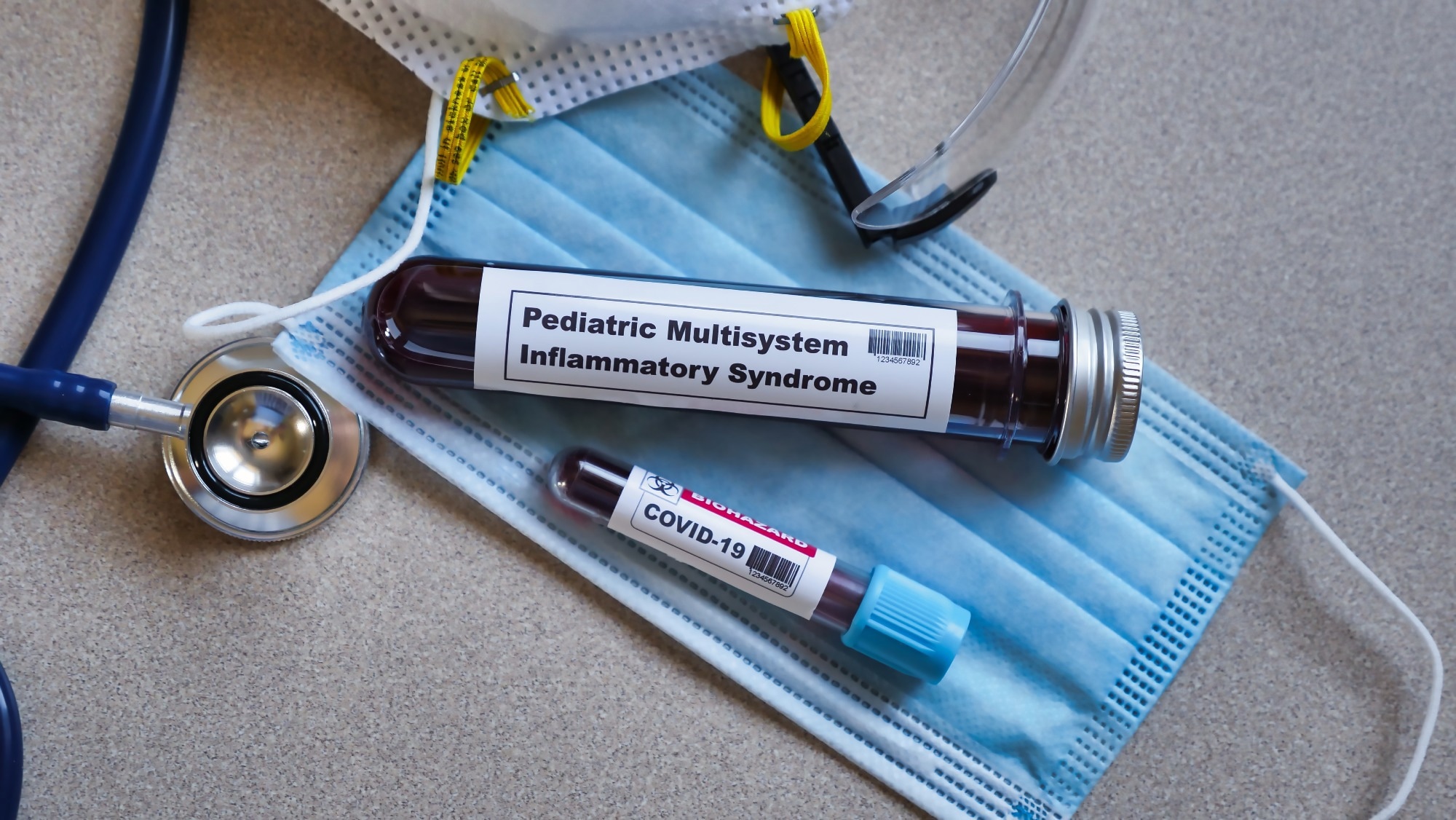
In a recent study published in Science, researchers assessed the inborn abnormalities of 2′-5′-oligoadenylate synthetase (OAS)-ribonuclease L (RNASEL) noted in coronavirus disease 2019 (COVID-19)-associated multisystem inflammatory syndrome in children (MIS-C).

In unvaccinated patients, the clinical variation in the course of severe acute respiratory syndrome coronavirus 2 (SARS-CoV-2) primary infection is enormous. Multisystem inflammatory syndrome in children (MIS-C), a severe phenotype associated with SARS-CoV-2, primarily affects children four weeks after infection. Most MIS-C cases are positive for anti-SARS-CoV-2 antibodies, while nearly all cases report previous SARS-CoV-2 exposure. MISC has numerous genetic, cellular, side effects moduretic tablets and clinical abnormalities, but the underlying cause is unknown.
About the study
In the present study, researchers described the autosomal recessive OAS1, OAS2, or RNASEL deficits among five unrelated children with MIS-C.
The team conducted whole-genome or whole-exome sequencing on 558 MIS-C patients from the COVID Human Genetic Effort (CHGE) international cohort. Homozygous or hemizygous variants predicted to be loss-of-function (pLOF) in human genes and had a gene damage index (GDI) of less than 13.83 were detected. Consequently, the search was expanded to include all homozygous or probable compound heterozygous nonsynonymous or important splicing site variants having a minor allele frequency (MAF) of less than 0.01 at four different loci in the MIS-C group. To examine the expression and role of these 16 variants in vitro, the team investigated RNA degradation mediated by RNase L in RNase L-deficient HeLa M cells cotransfected with the respective OAS1, OAS2, OAS3, or RNASEL complementary deoxyribonucleic acid (cDNA).
The team also studied whether mononuclear phagocytes deficient in OAS-RNase L had heightened inflammatory responses against SARS-CoV-2. THP-1 cells with knockouts (KO) of RNase L, OAS1, or OAS2, stimulated with either SARS-CoV-2 or intracellular poly(I:C), were subjected to bulk RNA sequencing.
Results
The study results discovered two unrelated patients who were homozygous for stop–gain mutations of OAS1 and RNASEL. OAS1 is among the four OAS family members, including OAS1, OAS2, OAS3, and OASL. These proteins constitute type I interferon (IFN)-inducible cytosolic proteins, which generate 2′-5′-linked oligoadenylate (2-5A) after binding to double-stranded (ds) RNA. In turn, the 2-5A triggers the dimerization and consequent stimulation of the latent endoribonuclease RNase L, which destroys viral or human single-stranded RNA (ssRNA).
Antibodies eBook

Even though RNase L, OAS1, OAS2, and OAS3 are expressed in various mouse and human cell types, their levels are notably high among myeloid cells such as macrophages and monocytes. Consequently, autosomal recessive (AR) abnormalities of the OAS-RNase L pathway may explain MIS-C by compromising the inhibition of viral replication and/or amplifying the inflammatory response in macrophages, monocytes, dendritic cells, or other types of cells triggered by viral exposure.
Consequently, autosomal recessive (AR) abnormalities of the OAS-RNase L pathway may explain MIS-C by compromising the inhibition of viral replication and/or amplifying the inflammatory response in macrophages, monocytes, dendritic cells, or other types of cells triggered by viral exposure. Furthermore, the team found 12 unrelated patients and 16 variants of RNASEL, OAS1, OAS2, and OAS3.
The three mutant OAS2 proteins identified, namely p.Q258L, p.R535Q, and p.V290I, were generated in normal proportions, but p.R535Q showed insignificant activity, and p.Q258L and p.V290I were less active than the wild-type (WT) protein. Except for p.R932Q, all OAS3 variants were generated in normal quantities and exhibited normal activity levels. The p.E265* variant of RNASEL was produced as a truncated protein and displayed LOF, while the p.I264V variant exhibited neutral expression and function. P1's OAS1 variant showed LOF, while P2, P3, and P4's OAS2 variants were hypomorphic.
Exogenous 2-5A, which is normally produced by OASs after dsRNA sensing and is responsible for RNase L activation, rescued the inflammatory phenotype of OAS1 KO THP-1 cells stimulated intracellularly with poly(I:C) following exogenous 2-5A treatment. Dephosphorylated 2-5A, which was incapable of activating RNase L, had no such impact. Additionally, exogenous 2- 5A administration inhibited the responsiveness of WT THP-1 cells to toll-like receptor 7 (TLR7)/8 activation. In RNase L-KO or -KDn THP-1 cells, 2-5A treatment produced a significantly reduced effect or no inhibitory effect at all.
Gene-set enrichment analysis (GSEA) showed enrichment among genes associated with inflammatory responses and IFN-gamma signaling in cells having OAS– RNase L deficiency, indicating that these cells exhibited a heightened inflammatory response against synthetic dsRNA as well as SARS-CoV-2. In addition, RNase L KO THP-1 cells secreted higher proportions of CXCL10 and interleukin (IL)-6 as compared to that of WT cells when they were co-cultured with SARS-CoV-2–infected Vero cells, which promote SARS-CoV-2 reproduction. These data showed that OAS-RNase L deficiency causes increased inflammatory responses among mononuclear phagocytes upon abortive SARS-CoV-2 infection as well as co-culture with cell types that replicated SARS-CoV-2.
Overall, the study findings indicated that human RNase L, OAS1, and OAS2 are required to properly regulate protection against SARS-CoV-2 but are essentially redundant under natural infection settings. It is also evident that the RNase L-dependent actions of OAS1 and OAS2 are essential for the modulation of immunity against SARS-CoV-2 within the same cells, as genetic loss of either of these three cell components leads to the same clinical and immunological phenotype, MIS-C.
- Lee, Danyel; Le Pen, Jérémie; Yatim, Ahmad; Dong, Beihua; Aquino, Yann; Ogishi, Masato; Pescarmona, Rémi; Talouarn, Estelle; Rinchai, Darawan; Zhang, Peng; Perret, Magali; et al. (2022). Inborn errors of OAS–RNase L in SARS-CoV-2–related multisystem inflammatory syndrome in children. Science. doi: https://doi.org/10.1126/science.abo3627 https://www.science.org/doi/10.1126/science.abo3627
Posted in: Medical Science News | Medical Research News | Disease/Infection News
Tags: Allele, Antibodies, Autosomal, Cell, Children, Compound, Coronavirus, Coronavirus Disease COVID-19, covid-19, CXCL10, Exome Sequencing, Frequency, Gene, Genes, Genetic, Genome, immunity, in vitro, Interferon, Interleukin, Intracellular, Phagocytes, Phenotype, Protein, Receptor, Reproduction, Respiratory, RNA, RNA Sequencing, SARS, SARS-CoV-2, Severe Acute Respiratory, Severe Acute Respiratory Syndrome, Splicing, Syndrome

Written by
Bhavana Kunkalikar
Bhavana Kunkalikar is a medical writer based in Goa, India. Her academic background is in Pharmaceutical sciences and she holds a Bachelor's degree in Pharmacy. Her educational background allowed her to foster an interest in anatomical and physiological sciences. Her college project work based on ‘The manifestations and causes of sickle cell anemia’ formed the stepping stone to a life-long fascination with human pathophysiology.
Source: Read Full Article
