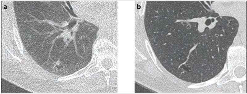
According to an article in ARRS’ American Journal of Roentgenology (AJR), Lung-RADS—as applied by radiologists in clinical practice—achieved excellent performance for lung cancer risk assessment on follow-up screening CT examinations, although strict application did downgrade some malignancies.
Noting that volumetric assessments showed weaker performance than clinical Lung-RADS, “new nodules warrant smaller size thresholds than existing nodules,” clarified coauthors Mark M. Hammer and Suzanne C. Byrne from Brigham and Women’s Hospital at Harvard Medical School in Boston, MA.
The pair’s retrospective study included 185 patients (100 women, 85 men; median age, 66 years) who underwent lung cancer screening CT examinations for which a prior CT was available. To enrich that sample with suspicious nodules, Hammer and Byrne performed stratified random sampling, which yielded 50, 45, 47, 30, and 13 nodules with Lung-RADS categories 2, 3, 4A, 4B, and 4X, respectively. Extracting nodule linear measurements from clinical reports to generate Lung-RADS categories via strict criteria, a semiautomated tool was used to obtain nodule volumes, which then generated NELSON categories.
With 29 cancers diagnosed, the weighted cancer risk was 5% for new nodules, 1% for stable existing nodules, and 44% for growing existing nodules. Whereas no clinical category 2 nodule was cancer, using strict Lung-RADS, 34 nodules, including 7 cancers, were downgraded to category 2. For clinical Lung-RADS categories, AUC was 0.96, compared with 0.71–0.84 for volumetric NELSON (two readers).
Source: Read Full Article
