What is fetal hemoglobin?
Fetal hemoglobin (HbF) is the most dominant form of hemoglobin (Hb) in fetuses and persists until birth, at which time the production of adult Hb is upregulated. Both fetal and adult Hb contain alpha (α) globin chains; however, in adult Hb, the gamma (γ) globin chains are replaced with beta (β) globin.
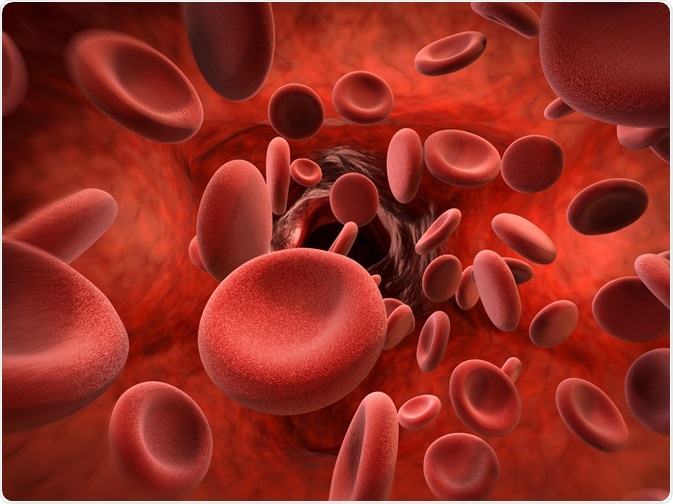
 Image Credit: Phonlamai Photo / Shutterstock.com
Six months after birth, HbA is the dominant form of Hb, and any conditions which affect Hb become clinically apparent. At six months, HbF comprises less than 5% of the total Hb, and it continues to fall, reaching the adult level of less than 1% by the age of 2.
The switch from fetal to adult Hb production does not occur to completion, nor is it irreversible. All adults retain the capacity to produce some HbF, which steadily declines throughout adult life. The degree to which HbF persists tends to vary greatly between adults, and this variability is under genetic control.
The persistence of relatively high levels of HbF production is not clinically harmful in healthy individuals; however, in patients suffering from conditions that affect the quality of HbA (such as those suffering from sickle cell disease or β-thalassemia), the condition can offer an advantage.
High HbF levels confer significant benefits – namely milder disease progression and fewer complications. Therefore, HbF has an ameliorating effect, which has prompted several approaches to therapeutic agent production in patients with mutations affecting adult HbA quality. These approaches include, for example, the pharmacological and gene transfer for HbF synthesis reactivation.
The balance between HbF and HbA
The residual HbF in adults is distributed unevenly amongst the red blood cells; cells that contain measurable quantities are referred to as F cells (FC). These typically contain a mean cell volume (MCV) of between 80-90 fl, compared to larger fetal cells with an MCV of approximately 125 fl. Within the FC, HbF only comprises a small portion of the total Hb.
Detection of FC occurs traditionally by an acid illusion method developed by Kleihauer; however, a much more sensitive immunofluorescence-based method uses an anti-γ globin antibody for use on fixed red blood cell smears, or via fluorescence-activated cell sorting (FACS), following labeling of the intracellular HbF.
Surveys of healthy blood donors across population groups demonstrate that levels of FC and HbF can vary more than 20-fold, and the distribution is both continuous and positively skewed. Furthermore, family studies show levels of HbF tend to be heritable, although the inheritance patterns are not clear. Twin studies show that the correlation between HbF and FC is genetically controlled, with a heritability of 0.89.
Patterns of inheritance
Hereditary persistence of fetal hemoglobin is a condition in which levels of HbF persist at levels greater than typically expected (less than 1%). In hereditary persistence of fetal hemoglobin (HPFH), this HbF percentage varies from levels as low as 0.8-1.0% to approximately 30% of the total hemoglobin.
However, the percentage of HbF can climb as high as 100% in homozygotes (individuals with two copies of the affected gene) for delta beta (δβ) thalassemia, another form of thalassemia characterized by defective mutations in the δβ chains of hemoglobin.
This condition is inherited in a mendelian fashion and is caused either by large deletions in the gene that controls the β subunit for HbA or by point mutations in the promoter (site of gene expression) of the γ globin genes. The condition is both benign and asymptomatic. The condition is classified as either pancellular / homocellular or heterocellular, based on the hemoglobin distribution pattern.
Pancellular conditions affect all cells, whereas heterocellular conditions affect some cells, and homocellular conditions affect similar groups of cells. In heterocellular HPFH, the level of HBF can range from 10 to 40%. On the other hand, the levels of HbF in heterocellular HPFH are only modestly elevated, and levels of hemoglobin are unevenly distributed amongst the red blood cells.
Pathogenic increases in HbF, which are distributed heterogeneously, are associated with drug use, malignancies, and hemoglobinopathies. Drugs such as hydroxyurea and pomalidomide increase HPFH. Trisomy 13, a chromosome disorder, is associated with a switch from HbF to HbA with persistently elevated HbF quantities in the blood.
Some patients with β-thalassemia, which is caused by reduced or absent synthesis of the β globin chains of hemoglobin which reduces the oxygen-carrying capacity of red blood cells, contain elevated levels of HbF that correlate with the degree of β chain deficiency as well as a co-inheritance of α thalassemia.
This protects against the deleterious effect of α- globin chain precipitation, caused by decreased α cell: γ cell chain ratios, which result in unstable tetramers, which are poor carriers of O2.
Other conditions in which elevated HbF is seen in patients include leukemia, following chemotherapy, as well as in patients treated for severe iron deficiency anemia due to acute blood loss. This occurs as a result of 'stressed' hematopoiesis or erythropoiesis (hemoglobin or red blood cell production, respectively). Patients with inherited bone marrow failure syndromes additionally show altered HbF as part of stressed synthesis blood components.
The benefits of HPFH are most noticeable when it co-exists with sickle cell or β thalassemia. These conditions cause diminished oxygen-carrying capacity of red blood cells due to unstable chains that form part of the tetramer of the oxygen-binding hemoglobin. As a result, the production of HbF alleviates this issue, as HbF contains γ rather than β chains, which are affected in adult Hb.
Owing to its ability to alleviate the severity of hemoglobinopathies in thalassemias, hereditary persistence of fetal hemoglobin tends to be selected for in populations in which these disorders are prevalent, such as people of African and Greek descent.
Sources
Friedman S, Schwartz. Hereditary persistence of foetal haemoglobin with beta-chain synthesis in cis position (gamma-beta+-HPFH) in a negro family". Nature. 1976 doi:10.1038/259138a0
Patel, S. et al. Inheritance of Hereditary Persistence of Fetal Haemoglobin (HPFH) in a Family of Western Odisha, India. J Clin Diagn Res. 2015. doi: 10.7860/JCDR/2015/12878.6548
Sokolova, A. et al. Hereditary persistence of hemoglobin F is protective against red cell sickling. A case report and brief review. Hematology/Oncology and Stem Cell Therapy. doi: 10.1016/j.hemonc.2017.09.003
Collins, F.S. et al. gamma beta+ hereditary persistence of fetal hemoglobin: cosmid cloning and identification of a specific mutation 5′ to the G gamma gene. Proc Natl Acad Sci USA. 1984. doi: 10.1073/pnas.81.15.4894
Thein, S. Discovering the genetics underlying foetal haemoglobin production in adults. British Journal of Haematology. 2009. doi: 10.1111/j.1365-2141.2009.07650.x
Further Reading
- All Hereditary Persistence of Fetal Hemoglobin Content
Last Updated: Jan 6, 2020

Written by
Hidaya Aliouche
Hidaya is a science communications enthusiast who has recently graduated and is embarking on a career in the science and medical copywriting. She has a B.Sc. in Biochemistry from The University of Manchester. She is passionate about writing and is particularly interested in microbiology, immunology, and biochemistry.
Source: Read Full Article
