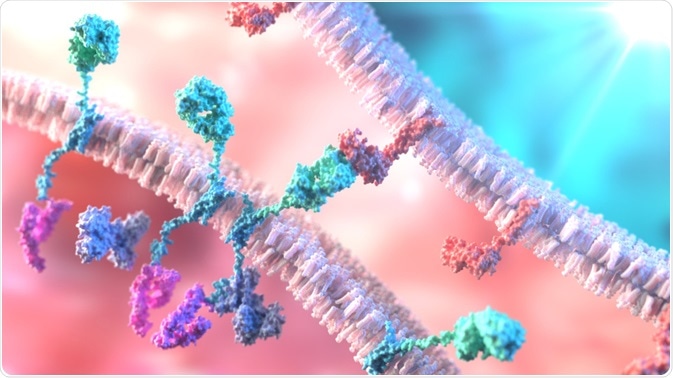
Chimeric antigen receptor (CAR) T-cell therapy is a recent promising therapeutic development for the treatment of various relapsing blood and bone marrow cancers, including lymphoma, myeloma, leukemia, and sarcoma.
Whilst the therapy itself has shown incredible therapeutic responses in patients, the side effects are significant and toxic. The two major adverse effects of CAR T-cell therapy are cytokine release syndrome (CRS) and neurotoxicity.

Image Credit: Alpha Tauri 3D Graphics/Shutterstock.com
What is Cytokine Release Syndrome?
CRS is characterized by fever, low blood pressure (hypotension), low tissue oxygenation (hypoxia) and some degree of multi-system organ failure. CRS can happen in almost every patient undergoing CAR T-cell therapy and tends to happen almost immediately after therapy.
However, it can be managed by treating patients with specific anti-inflammatory drugs, such as the IL-6 inhibitor, tocilizumab, without adversely affecting the CAR T-cell therapy itself.
Neurotoxicity in CAR T-Cell Therapy
With respect to the neurotoxicity of CAR T-cell therapy, the mechanisms underlying this remain poorly understood and the clinical presentations of the neurological side effects are variable between patients.
Symptoms usually begin after a short delay and do not present in all patients. Some of the common neurotoxic effects include headache, cerebral oedema, delirium (mental confusion), general weakness, cognitive changes, encephalopathy (brain-damaging disease), seizures, tremors and ataxia (uncoordinated balance).
Unfortunately, medications used for CRS do not typically have any beneficial effect on the neurotoxic side effects or symptoms.
Neurotoxicity in CAR T-cell therapy leads to increased cerebrospinal fluid (CSF) protein and white blood cell counts.
Patients with neurotoxicity have altered T-cell migration into the central nervous system, coupled with very high levels of interferon-gamma, interleukin-6 and tumor necrosis factor-alpha, which all indicate the breakdown of the blood-brain-barrier allowing T-cells and cytokines into the CSF, as well as increased production of cytokines within the nervous system.
Neuroimaging studies, using MRI, have revealed that patients with neurotoxicity have patchy T2 hyperintensities in the white matter of the brain, as well as in some deep grey matter structures such as the thalamus.
These hyperintensities are indicative of cerebral oedema and could be due to small microscopic bleeding occurring within the brain. Electroencephalograph (EEG) monitoring has revealed diffuse slowing EEG patterns that resemble patterns in critically ill patients with encephalopathy.
Neurotoxicity only occurs in patients who also have developed CRS with CAR T-cell therapy. However, the neurotoxicity in CAR T-cell therapy presents later than CRS and takes much longer to resolve. It does not typically respond to IL-6 treatment that improves CRS.
Although it is not known exactly how neurotoxicity occurs, it is known that it occurs in people who may have pre-existing neurological issues, have a higher tumor burden, have higher CAR T-cell dosage, or receive fludarabine for cancer treatment.
Some studies have shown that key cytokines may be important in the development of neurotoxicity. Within the first 36 hours after CAR T-cell therapy, patients who develop the most severe neurotoxicity usually have higher serum levels of MCP-1, IL-15, IL-10, and IL-2 as well as much earlier peak of IL-6 compared to those with less severe neurotoxicity.
Thus, the prediction of severe neurotoxicity using these cytokines as biomarkers may be an effective predictive tool using serum isolated from patients within 36 hours after therapy, although providing this information in real-time using current technology is not viable, as most assays require time.
Management of Neurotoxicity
Most medications used to treat CRS do not have any beneficial therapeutic effect on neurotoxicity. In most cases where neurotoxicity occurs, the condition usually resolves with only supportive care within 21 days after CAR T-cell therapy, however, a very small percentage of cases may be fatal.
CRS can be managed effectively with tocilizumab (IL6 antagonist), and many caregivers only use supportive care to manage symptoms, though some suggest an early and aggressive treatment.
For those that opt for immunomodulatory treatments for neurotoxicity, tocilizumab is not often used to its limited therapeutic effects on neurotoxicity, but the overall very effective response for CRS. Corticosteroids, such as dexamethasone, can be used to treat cerebral oedema.
However, corticosteroids may reduce the effectiveness of CAR T-cell therapy in the treatment of cancers. However, there is some evidence that short courses of low dosage dexamethasone (10mg twice daily: 2-4 doses) does not seem to interfere with T-cell therapeutic effects.
However, high dosages of strong corticosteroids such as methylprednisolone at 1g/day can significantly reduce circulating T-cell counts.
More recently, siltuximab; a monoclonal antibody against IL-6, has shown evidence of efficacy against CRS and neurotoxicity without interfering with CAR T-cell therapy itself. However, it has not been approved by regulatory bodies for this purpose. Plasma exchange and platelet hypertransfusion may also be beneficial, although more research is needed to support their efficacy.
In summary, there are two main side effects of CAR T-cell therapy uses in the treatment of malignant cancers: cytokine release syndrome (CRS), which affects many recipients, and neurotoxicity, which affects a small subset of people with CRS. Drugs used to treat CRS, such as tocilizumab, do not typically have any therapeutic effect in neurotoxic side effects, which include headache, cerebral oedema, seizures, encephalopathy, and tremors.
Specific low-dosage corticosteroids may be able to treat and manage neurotoxicity without altering T-cell counts, though the mechanisms underlying neurotoxicity and its treatment are still poorly understood.
Sources:
- Rubin et al, 2019. Neurological Toxicities Associated With Chimeric Antigen Receptor T-cell Therapy. Brain 142 (5), 1334-1348. pubmed.ncbi.nlm.nih.gov/…/
- Gust et al, 2018. Neurotoxicity Associated With CD19-Targeted CAR-T Cell Therapies. CNS Drugs 32 (12), 1091-1101. pubmed.ncbi.nlm.nih.gov/…/
- Yanez et al, 2019. CAR T Cell Toxicity: Current Management and Future Directions. Hemasphere 3 (2), e186. pubmed.ncbi.nlm.nih.gov/…/
Further Reading
- All Therapeutics Content
Last Updated: Apr 10, 2020

Written by
Osman Shabir
Osman is a Neuroscience PhD Research Student at the University of Sheffield studying the impact of cardiovascular disease and Alzheimer's disease on neurovascular coupling using pre-clinical models and neuroimaging techniques.
Source: Read Full Article
