
One of the most exciting things to look forward to when a new baby is about to be born is the question, will it be a girl or a boy? While many and astonishingly diverse answers have been offered to this query, the use of modern ultrasonography offers an accurate answer from early pregnancy onwards.
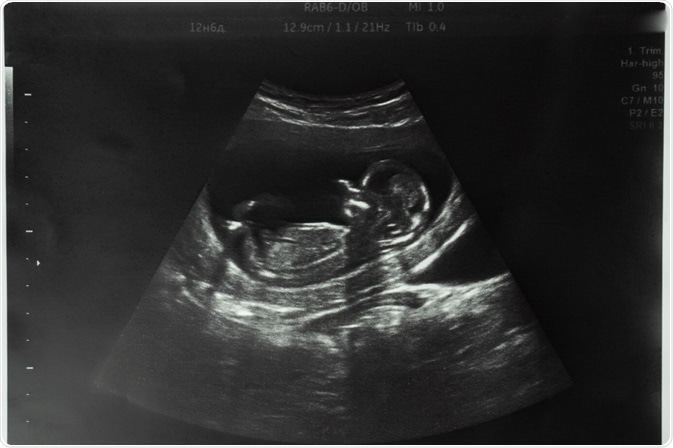
Image Credit: SViktoria/Shutterstock.com
Prenatal Sex Diagnosis by biopsy
The prenatal diagnosis of sex is important in X-linked disorders such as Duchenne muscular dystrophy (DMD). The first step is via chorionic villus sampling (CVS), at 11-13 weeks to avoid limb reduction defects. Here, a few of the placental villi are removed for genetic analysis.
This admittedly risky procedure may lead to miscarriage in 0.5-1% of cases, especially when carried out in centers with less experience in this procedure as assessed by the number of cases each year.
Amniocentesis, the aspiration of a sample of amniotic fluid to analyze the DNA from the cells in it, is another procedure used for sex determination, but due to the risk of talipes equinovarus, it is done only after 15 weeks of gestation. Compared to the former technique, the learning curve is surmounted in 30 procedures vs 400 for CVS. Moreover, CVS sampling failure occurs at a three-fold higher level relative to amniocentesis.
Non-Invasive Sex Determination
It was in 1997 that cell-free fetal genetic material (deoxyribonucleic acid, DNA), called cff-DNA, was first detected in maternal plasma. This allowed scientists to think about using this blood test for non-invasive prenatal testing (NIPT) of sex and genetic disorders.
In maternal serum, cff-DNA is found only after 7 weeks of pregnancy but vanishes from two hours following delivery. It includes placental DNA, as well as somatic fetal DNA. Overall, it makes up to 6% of the total cell-free DNA to be found in the mother’s blood during early and late pregnancy.
To determine the sex of the fetus, cff-DNA is analyzed using polymerase chain reaction (PCR) to identify sequences found only on the Y chromosome. This can be done in the period before 9 weeks, after which ultrasound examination can be done with a high degree of accuracy.
The benefits of cff-DNA are the non-invasive sampling method, absence of miscarriage risk, safety to the mother, and the ability to determine the fetal sex from 7 weeks onwards, and thus earlier than 11-13 weeks, which is when CVS is ideally done.
Both false positives, from a vanishing twin or placental mosaicism, or simply contamination, and false negatives, because the Y chromosome sequence fails to be detected, are possible. Further refinement of the technique is still underway.
Ultrasound sex determination
High-resolution ultrasound now allows fetal morphology to be visualized clearly, thus paving the way for fetal sex determination at an earlier date during the first trimester. At an embryonic length (crown-rump length, CRL) of 76 mm or more, the chances of identifying the fetal sex accurately are 99% or more.
Early in week 5 of pregnancy, a pair of cloacal folds form in the future genital area, joining to form the midline genital tubercle. This will form the penis or clitoris but looks the same until about 9 weeks (CRL ~33 mm).
Various ultrasonic criteria have been proposed, such as the ‘dome sign’, and the ‘sagittal sign’ (the caudal or cranial direction of the genital tubercle relative to a horizontal line through the lumbosacral skin surface, respectively).
The sagittal sign can achieve 99-100% accuracy, though in about one in seven diagnosis is not possible due to intermediate-facing positions of the genital tubercle or unfavorable fetal position. The feasibility of prediction goes up from about 40% at 50 mm to 97% at 60 mm and above.
The chances of successful prediction are highest with the CRL above 66 mm (~13 weeks of gestation) and are low below 50 mm (~11+4 weeks of gestation). This corresponds to 9 weeks of gestation, which is when male-female differentiation begins.
Between 50-55 mm, that is, 11+4 weeks to 12 weeks of gestation, the accuracy is about 75%, but jumps to 96% with a CRL of 55-60 mm. For male fetuses, in fact, it is 99% vs 93.5% in females at this time. As stated above, by 12+2 weeks, with a CRL of 60 mm or more, the accuracy is 100% for both sexes.
Males are more likely to receive an accurate identification than females, below a length of 65 mm. Sonographic experience does not, thankfully, seem to play a part, with one study claiming that one week of experience is sufficient to achieve an accurate diagnosis.
Other ultrasound findings that have been discussed in terms of sex determination include the identification of the fetal scrotum, the midline raphe of the penis, the labia, the uterus, the testes within the scrotum, and the urination trajectory and origin in boys.
Does Maternal Intuition Work?
An interesting study has demonstrated that while mothers often feel they “know” the sex of their babies before birth, the predictions were correct only about half the time.
What Is the Conclusion?
Fetal gender may reliably be determined when CRL ≥ 60 mm (gestational age ≥ 12+2). Male gender may already be reliably determined when CRL ≥ 55 mm (gestational age ≥ 12+0). If CRL < 50 mm (gestational age < 11+4) the gender cannot be reliably predicted.”
References:
- Bowman-Smart, H. et al. (2019). Sex selection and non-invasive prenatal testing: A review of current practices, evidence, and ethical issues. Prenatal Diagnosis. https://doi.org/10.1002/pd.5555. https://obgyn.onlinelibrary.wiley.com/doi/full/10.1002/pd.5555
- Savirón-Cornudella, R. et al. (2021). Ultrasound measurement learning of fetal sex during the first trimester: does the experience matter?https://doi.org/10.2147/RRFU.S88738. https://www.dovepress.com/ultrasound-measurement-learning-of-fetal-sex-during-the-first-trimeste-peer-reviewed-fulltext-article-RRFU
- Becker, S. et al. (2004). Fetal gender and sonographic assessment of crown–rump length: implications for multifetal pregnancy reduction. Ultrasound in Obstetrics and Gynecology. https://doi.org/10.1002/uog.1084. https://obgyn.onlinelibrary.wiley.com/doi/full/10.1002/uog.1084
- Lubusky, M. et al. (2012). Ultrasound evaluation of fetal gender at 12-14 weeks. Biomedical papers of the Medical Faculty of the University Palacký, Olomouc, Czechoslovakia. doi: 10.5507/bp.2012.022. Epub 2012 Apr 19. https://pubmed.ncbi.nlm.nih.gov/22660228/
- Odeh, M. et al. (2021). Sonographic fetal sex determination. Obstetric and Gynecologic Survey. doi: 10.1097/OGX.0b013e318193299b. https://pubmed.ncbi.nlm.nih.gov/19099612/
- McFadzen, M. et al. (2017). Maternal Intuition of Fetal Gender. Journal of Patient-Centered Research and Reviews. https://doi.org/10.17294/2330-0698.1454. https://pubmed.ncbi.nlm.nih.gov/31413978/
- Miura, K. et al. (2011). Clinical application of fetal sex determination using cell-free fetal DNA in pregnant carriers of X-linked genetic disorders. Journal of Human Genetics. https://doi.org/10.1038/jhg.2011.7. https://www.nature.com/articles/jhg20117
Further Reading
- All Pregnancy Content
- Early Signs of Pregnancy
- Is it Safe to Exercise During Pregnancy?
- Pregnancy: 0-8 weeks
- Pregnancy: 9 – 12 weeks
Last Updated: Sep 20, 2021

Written by
Dr. Liji Thomas
Dr. Liji Thomas is an OB-GYN, who graduated from the Government Medical College, University of Calicut, Kerala, in 2001. Liji practiced as a full-time consultant in obstetrics/gynecology in a private hospital for a few years following her graduation. She has counseled hundreds of patients facing issues from pregnancy-related problems and infertility, and has been in charge of over 2,000 deliveries, striving always to achieve a normal delivery rather than operative.
Source: Read Full Article