
In a recent study posted to the bioRxiv* preprint server, researchers proposed an optimal system to generate severe acute respiratory syndrome coronavirus 2 (SARS-CoV-2) virus-like particles (VLPs) for examining viral assembly and entry into target host cells.
Background
VLPs are non-infectious multi-protein-lipid structures that mimic the conformation of a native virus and could be used to produce efficient and cheap vaccine candidates for coronavirus disease 2019 (COVID-19). Moreover, non-infectious VLPs have allowed examining the SARS-CoV-2 entry and assembly in biosafety level 2 (BSL-2) facilities.
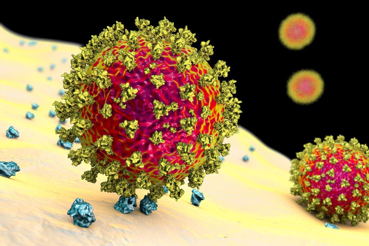
SARS-CoV-2, ondansetron aman buat ibu hamil the causative agent of COVID-19, is a ribonucleic acid (RNA) enveloped virus composed of 29 structural proteins. The four main structural proteins – membrane (M), nucleocapsid (N), envelope (E), and spike (S) proteins assemble to form 100nm SARS-CoV-2 particles.
Several reports have suggested that the different combinations of the M, N, E, and S proteins, play a crucial role in VLPs formation. Researchers have established several minimal systems for SARS-CoV-2 VLPs production; however, to date, an optimal proportion of SARS-CoV-2 structural proteins for use in such systems has not been determined and tested.
About the study
In the present study, researchers used an optimal transfected plasmid ratio of M, N, E, and S proteins mimicking SARS-CoV-2 RNA ratios to obtain SARS-CoV-2 VLPs composed of structural proteins M, N, E, and S.
They derived SARS-CoV-2 RNA ratios from the transcriptomic analysis of the SARS-CoV-2 genome in infected pulmonary Calu3/intestinal Caco2 cells and human pulmonary A549hACE2 cells. They also optimized the production of fluorescent VLPs (bi- or mono-color) tagged with structural SARS-CoV-2 proteins to visualize viral particle entry into host cells using quantitative confocal microscopy.
Further, they characterized these VLPs using advanced microscopic techniques to determine the numbers, size, and morphology of VLPs comparable to wild-type (wt) SARS-CoV-2 particles. They used a suite of microscopic techniques, including Fluctuation Correlation Spectroscopy (FCS), Total Internal Reflection Fluorescence Microscopy (TIRF-M), Photo-Activable Localization Microscopy (PALM), and correlative fluorescence-Atomic Force Microscopy (AFM) for the analysis.
Study findings
RNA sequencing (RNAseq) analysis showed that a plasmid ratio of 3:12:2:5 for M:N: E:S increased VLPs production by 1.5-fold for VLPs composed of MNE and MNES. Comparatively, the 3:3:3:5 ratio for M:N: E:S with more S incorporation and more mature S2 on the VLPs, mimicked the wt virus.
The AFM results confirmed that these viral particles were VLPs and not exosomes or extracellular vesicles (EVs). Immunospotting on green fluorescent protein (GFP)-VLP showed less than 5% or no colocalization between VLP and EVs tagged with a cluster of differentiation 81 (CD81) marker, suggesting VLPs exited through a distinct secretory pathway.
The authors measured MNES VLPs of size 126 ± 17 nm using AFM. They produced VLPs composed of M or N proteins in fusion with fluorescent proteins; intriguingly, not only did SARS-CoV-2 VLPs tolerate the incorporation of fluorescent M and N tagged proteins, this tagging did not temper the viral particle assembly. This helped researchers establish the incorporation of the mature S in fluorescent VLPs. Then, using FCS and PALM, they found a particle size range of 100-110 nm for MNE VLPs, and 130nm for MNES VLPs, consistent with the AFM results.
The FCS results showed two subsets of the MNE and MNES VLP sizes: one bigger than 100nm and one equal to 100nm in diameter, suggesting that SARS-CoV-2 produced some particles with and without S, as observed by immunospotting as well.
The immunospotting results confirmed that at least 25% of the M(GFP)NES VLPs incorporated the S protein. According to the authors, it would be interesting to vary the ratio of S expression in the experimental cells to see if it would change the level of S incorporation in the VLPs. Likewise, incorporating MNES VLP mimicking the SARS-CoV-2 into animal models could provide sufficient amounts of neutralizing antibody responses without being infectious.
Lastly, at the human pulmonary cell surface, the M(GFP)NES VLPs could recognize the human angiotensin-converting enzyme 2 (hACE2) receptor and endocytosed, confirming that the S incorporated in the VLPs remained functional.
Conclusions
Overall, the study findings reaffirmed that the non-fluorescent and fluorescent VLPs composed of the SARS-CoV-2 M, N, E, and S proteins could package SARS-CoV-2 RNA to transduce and express genes in the target host cells.
Furthermore, their optimal production and detailed characterization could help understand the underlying mechanisms of the SARS-CoV-2 assembly and host cell entry in a safe environment (BSL-2 and not BSL-3) and even aid in the development of VLP-based COVID-19 vaccines.
*Important notice
bioRxiv publishes preliminary scientific reports that are not peer-reviewed and, therefore, should not be regarded as conclusive, guide clinical practice/health-related behavior, or treated as established information.
- Gourdelier, M. et al. (2022) "Optimized production and fluorescent labelling of SARS-CoV-2 Virus-Like-Particles to study virus assembly and entry". bioRxiv. doi: 10.1101/2022.03.23.485575. https://www.biorxiv.org/content/10.1101/2022.03.23.485575v1
Posted in: Medical Science News | Medical Research News | Disease/Infection News
Tags: Angiotensin, Angiotensin-Converting Enzyme 2, Antibody, Atomic Force Microscopy, Cell, Confocal microscopy, Coronavirus, Coronavirus Disease COVID-19, covid-19, Enzyme, Exosomes, Fluorescence, Fluorescence Microscopy, Fluorescent Protein, Genes, Genome, Localization Microscopy, Membrane, Microscopy, Morphology, Particle Size, Plasmid, Protein, Receptor, Respiratory, Ribonucleic Acid, RNA, RNA Sequencing, SARS, SARS-CoV-2, Severe Acute Respiratory, Severe Acute Respiratory Syndrome, Spectroscopy, Syndrome, Vaccine, Virus

Written by
Neha Mathur
Neha is a digital marketing professional based in Gurugram, India. She has a Master’s degree from the University of Rajasthan with a specialization in Biotechnology in 2008. She has experience in pre-clinical research as part of her research project in The Department of Toxicology at the prestigious Central Drug Research Institute (CDRI), Lucknow, India. She also holds a certification in C++ programming.
Source: Read Full Article