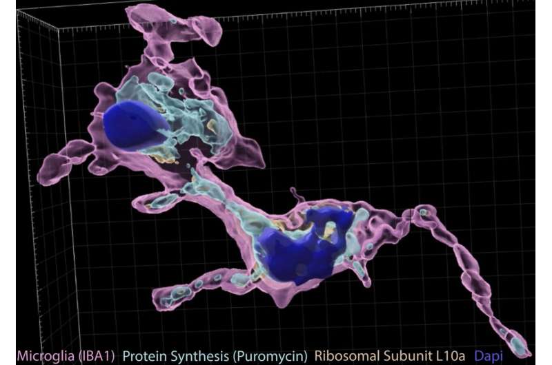
Recent studies have found that some cells of the brain, including neurons, astrocytes, and oligodendrocytes, can transport pieces of the genetic code (RNA) from the nucleus out to their distal processes which may be several microns away, and then locally translate proteins from this code within the process itself. Yet whether microglia, zoloft itching all over immune cells that protect the brain from damage and disease, might be able to engage in this process remains poorly understood.
Researchers at Washington University School of Medicine recently carried out a study exploring if microglial processes can support the local translation of protein. Their findings, published in Nature Neuroscience, suggest that microglia create new proteins within their peripheral processes, particularly at sites where they are “ingesting” (i.e., phagocytosing) cells or other targets that are no longer needed in the body.
“When you think about how complex the shapes of brain cells are, you realize they have this fundamental problem when it comes to gene expression,” Joseph D. Dougherty, one of the researchers who carried out the study, told Medical Xpress. “Think of a neuron with its hundreds of little processes and synapses: to strengthen a given synapse after activity (i.e., to help encode a new memory), the cell needs new proteins at just that synapse—but not everywhere else. Yet, the genes that code for these proteins pretty much reside in just one place that is far from the synapse, specifically in the nuclear DNA.”
When cells create new protein, this protein is delivered to target locations inside the cell via a sophisticated translational process. Firstly, DNA (i.e., the material carrying genetic information) is transcribed to the nucleic acid RNA, which is then translated into protein.
“Over the last couple of decades, researchers realized that neurons have the ability to localize where that RNA is translated by moving mRNA and the translational machinery (ribosomes) to the activated synapses,” Dougherty explained. “It is sort of like shipping the plans and the factory to where you need a lot of new parts. While neurons are the most famous cells in the nervous system, if you look at other cells like astrocytes and microglia, they have the same challenge of a complex morphology with lots of processes that may need to act independently. ”
Microglia are tiny and yet functionally refined immune cells that protect the brain and the body from infections, diseases and traumatic injuries. While many studies have investigated the functions and processes of these vital immune cells, their ability to locally translate protein within their processes has rarely been explored before.
“As the resident immune cells, microglia are both the gardeners and the watchdogs of the brain, with 5–10 little processes that are continuously moving and surveying the surrounding cells, occasionally pruning excess synapses that are no longer needed and clearing any debris from injury or infection by a process called phagocytosis (i.e., surrounding and ‘eating’ debris and unwanted cells),” Dougherty said. “Our key question was does this phagocytosis, like strengthening a synapse in a neuron, also need new protein production? And if so, how might a process on one side of the cell turn on phagocytosis while the other processes are still in surveillance mode?”
The key hypothesis put forward by Dougherty and his colleagues is that just like neurons and astrocytes, microglia can also locally translate protein, as this allows them to act independently from other cells, performing their main functions while also translating protein if needed. To confirm this hypothesis, they would need to gather three key observations.
First, if their suspicious were true, microscopic examinations would unveil instructions for producing new proteins, known as mRNA, among microglial processes. Among these same processes, they should have also been able to detect ribosomes. Finally, after examining brain slices treated with specific compounds that highlight new protein translation, the researchers should find evidence of newly translated protein within the microglial processes.
“We also wanted to know what they were actually making out there,” Dougherty said. “We thus adapted some old biochemical methods from prior decades that were used to purify different fragments from ground up mouse brains by their size/density to enrich for processes of all cell types, and then paired this with a genetic trick to add a tag to ribosomes only in microglia. ”
Dougherty and his colleagues only pulled ribosomes from microglial processes (which they had tagged using genetic tools) and sequenced the mRNAs they found on them. This allowed the to determine what proteins they were producing locally.
Interestingly, the researchers found that much of the genes translated by microglial processes were involved in phagocytosis (i.e., the “ingestion” of debris and unwanted cells). Overall, thus suggests that the local translation of protein is a necessary step for phagocytosis.
“We asked whether blocking translation with classic ribosome-blocking drugs could block phagocytosis in brain slices, and it could,” Dougherty said. “While this told us translation was important, it didn’t tell us where it was important. My partner Mike Vasek had noticed that sometimes when we made these brain slices, we accidently cut off just one process of a microglia. We started wondering if these were inert, or more like ‘The Thing’ from the Addam’s family, where maybe just this one process, detached from a nucleus, still conducted phagocytosis by itself?”
Dougherty, Vasek and their colleagues then conducted a final experiment to determine what would happen if they blocked protein translation. Their findings confirmed that microglial processes could act independently to engulf debris, and the translation of new protein allows them to do this most efficiently.
“Microglial processes are constantly moving—contacting hundreds of neighboring synapses and cells every hour,” Vasek, co-author of the paper, said. “They also do many different types of actions at their various contacts including pruning synapses, remodeling extracellular matrix, and phagocytosing an apoptotic cell. We found that microglia are making new protein within their peripheral processes and are making lots of new protein locally at sites where they are phagocytosing apoptotic cells and other targets.
“One exciting implication for our work is that it offers a possible explanation for how individual microglia can make proteins to execute a specific function in one distal part of its cell while making other subsets of proteins in other subcellular regions involved in other local tasks.”
The results gathered by this team of researchers offer new highly valuable insight into the complexity of microglial processes, while also confirming their ability to locally translate proteins without relying on protein “shipments” from the cell soma or center. In the future, they could pave the way for more research exploring microglial peripheral processes and protein translation, which could lead to more interesting discoveries.
“Now that we’ve established this happens, we are very interested in how they do this,” Dougherty added. “How do they localize the RNAs and ribosomes they need to the right location? What signals turn on local translation when they sense debris? How fine-tuned is this process? And does this process break down in diseases of the brain? We are excited to pursue these questions and many others with the new toolkit we devised.”
More information:
Michael J. Vasek et al, Local translation in microglial processes is required for efficient phagocytosis, Nature Neuroscience (2023). DOI: 10.1038/s41593-023-01353-0
Journal information:
Nature Neuroscience
Source: Read Full Article
