
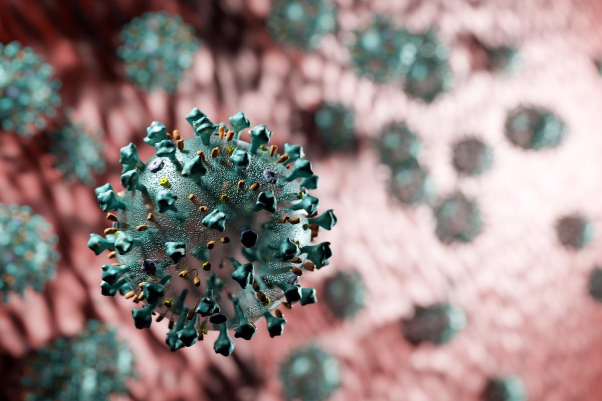
A recent study posted to the bioRxiv* preprint server investigated the susceptibility of macrophages to severe acute respiratory syndrome coronavirus 2 (SARS-CoV-2) virions.
Previous studies have characterized the replication cycle of SARS-CoV-2 primarily in cell lines and epithelial cells. However, the different representations of this cycle in other susceptible cell types, like the immune cells, require extensive research.
About the study
The present study assessed the sensitivity of macrophages towards infectious SARS-CoV-2 virions, causing the release of an anti-viral mediator and inflammatory response.
To model the differentiation of monocytes into macrophages, the team incubated human monocyte-derived macrophages (HMDM) with SARS-CoV-2 to quantify cytokine release and monitor macrophage responses. The team then investigated the susceptibility of HMDMs towards infection and replication of SARS-CoV-2 virions with SARS-CoV-2-permissive cultured human airway epithelial cells (Calu-3) considered as a positive control.
The authors then assessed the differences between the macrophages found in the lungs and HMDM, promethazine codeine 6.25 10 mg concerning their vulnerability to SARS-CoV-2 infection and replication. Airway macrophages were obtained from three donors and injected with SARS-CoV-2. Furthermore, the authors investigated the role of angiotensin-converting enzyme 2 (ACE2) expression in the resistance of macrophages against SARS-CoV-2 infection. This was assessed by evaluating the protein and messenger ribonucleic acid (mRNA) levels of ACE2 via quantitative polymerase chain reaction (qPCR) and immunoblotting.
The study also investigated the ability of SARS-CoV-2 to bind with and penetrate the HMDM surface, without the intervention of ACE2. Transmission electron microscopy determined the subcellular location of approaching virions.
Results
The study results showed that SARS-CoV-2 infection did not trigger the release of C–X–C motif chemokine 10 (CXCL10), interleukin 6 (IL-6), or tumor necrosis factor (TNF) in vitro. However, a robust secretion of TNF and IL-6 was observed in the presence of synthetic viral mimetics like the toll-like receptor (TLR) agonist, R848, and of CXCL10 for the melanoma differentiation-associated protein 5 (MDA5) ligand. Moreover, mRNA analyses showed that there was no upregulation of HMDM in the presence of SARS-CoV-2 virions while R848 and transfected poly I:C (pIC) caused high secretion.
The Calu3 cells were observed to be highly susceptible to SARS-CoV-2 at low as well as high dose infection as indicated by the growth of cell-associated viral RNA. However, these viral RNA levels showed no increase in the presence of infected-HMDM. Also, the newly synthesized viral protein was found in Calu3 control cells, with the expression of viral nucleoprotein (NP) increasing from two hours to 72 hours in Calu3. In contrast, HMDM did not have a newly synthesized viral NP as NP expression was barely detected 24 hours after infection. Overall, these findings showed that HMDM cells were not SARS-CoV-2-susceptible and did not support the generation of newly produced viral RNA and protein.
Furthermore, SARS-CoV-2-infected airway macrophages had no rise in cell-associated viral RNA between two to 24 hours of infection. However, the Vero E6 supernatant showed an increase in the levels of viral RNA which indicated productive viral replication and synthesis of viral RNA from infected cells, which was absent in the infected airway macrophages. This data highlighted that airway macrophages were not permissive to early viral replication stages.
Assessment of ACE2 protein and mRNA levels in HMDM showed low levels of ACE2 mRNA expression while no expression of ACE2 proteins was observed. Also, no ACE2 protein was found in airway macrophages. However, overexpression of ACE2 in THP-1 cells produced detectable levels of ACE2 protein and mRNA. The team also found detectable levels of transmembrane serine protease 2 (TMPRSS2) in THP-1-ACE2 and HMDM cells.
The study also showed that HMDM and THP-1-ACE2 had internalized virions that were intact in the phagosomal system while only the THP-1-ACE2 cells had virions that were bound and almost fused to the plasma membrane. These particular virions had a morphological structure that was similar to the new virions that were released from Calu3 cells.
The endogenous expression of TMPRSS2 suggested that the THP-1-ACE2 cells allow fusion of the virus with the plasma membrane, thus delivering the viral NP and genome into the cytoplasm. Altogether, these results show that even though HMDM allowed the uptake of SARS-CoV-2 into the phagosomal compartments, the low levels of ACE2 expression prevented the processing of the SARS-CoV-2 spike (S) protein and the fusion of the virus-cell membrane.
Conclusion
The study findings showed that the human macrophages are not susceptible to SARS-CoV-2 infections unless there is an increase in ACE2 expression and the early stage of viral replication begins. The researchers believe that this study provides a better insight into SARS-CoV-2 cell tropism and its impact on immune pathways. Furthermore, this study can help develop novel therapeutic targets for new medications to treat SARS-CoV-2 infections.
*Important notice
bioRxiv publishes preliminary scientific reports that are not peer-reviewed and, therefore, should not be regarded as conclusive, guide clinical practice/health-related behavior, or treated as established information.
- Larisa I Labzin, et al. (2022). ACE2 is necessary for SARS-CoV-2 infection and sensing by macrophages but not sufficient for productive viral replication. bioRxiv. doi: https://doi.org/10.1101/2022.03.22.485248 https://www.biorxiv.org/content/10.1101/2022.03.22.485248v1
Posted in: Medical Science News | Medical Condition News | Disease/Infection News
Tags: ACE2, Angiotensin, Angiotensin-Converting Enzyme 2, Cell, Cell Membrane, Chemokine, Coronavirus, Coronavirus Disease COVID-19, CXCL10, Cytokine, Cytoplasm, Electron, Electron Microscopy, Enzyme, Genome, in vitro, Interleukin, Ligand, Lungs, Macrophage, Melanoma, Membrane, Microscopy, Monocyte, Necrosis, Polymerase, Polymerase Chain Reaction, Protein, Receptor, Research, Respiratory, Ribonucleic Acid, RNA, SARS, SARS-CoV-2, Serine, Severe Acute Respiratory, Severe Acute Respiratory Syndrome, Syndrome, Tumor, Tumor Necrosis Factor, Virus

Written by
Bhavana Kunkalikar
Bhavana Kunkalikar is a medical writer based in Goa, India. Her academic background is in Pharmaceutical sciences and she holds a Bachelor's degree in Pharmacy. Her educational background allowed her to foster an interest in anatomical and physiological sciences. Her college project work based on ‘The manifestations and causes of sickle cell anemia’ formed the stepping stone to a life-long fascination with human pathophysiology.
Source: Read Full Article