The hippocampus is a key region of the mammalian brain, which has been found to be involved in the formation of episodic memories. These are long-term memories of events or experiences that can be consciously remembered and mentally re-experienced, such as recollections of a particularly memorable birthday party, a holiday and so on.
Essentially, the hippocampus is known to support the “conversion” of our life experiences into episodic memories that we can consciously recall for a long time. Past neuroscience studies found that this memory consolidation process, as well as the ability to learn from past experiences, is influenced by the generation of electrophysiological oscillations in the hippocampus, known as sharp-wave ripples (SWRs).
Researchers at Columbia University recently set out to better understand how specific populations of interneurons (i.e., navy medicine history local neurons that generally inhibit excitatory pyramidal neurons in cortical structures) regulate memory consolidation and learning while animals are actively engaged in activities. Their paper, published in Nature Neuroscience, characterizes the activity of interneurons in two regions of the hippocampus as mice move in their surrounding environment and their brain is forming new memories.
“After memories form, they need to be consolidated or they will vanish,” Tristan Geiller, one of the researchers who carried out the study, told Medical Xpress. “There is overwhelming evidence that memory consolidation occurs during events that are hundreds of milliseconds long, called SWR. One the biggest and long-standing questions in our field relates to identifying what triggers these SWR events. Over the years, inhibitory interneurons have been posited as a major circuit element supporting the generation of SWR, but it is notoriously difficult to experimentally record the activity of these cells in awake behaving animals and test this hypothesis.”
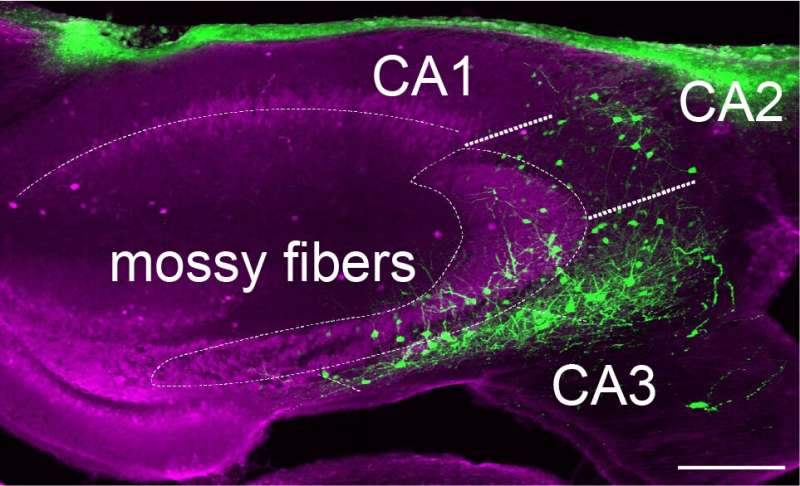
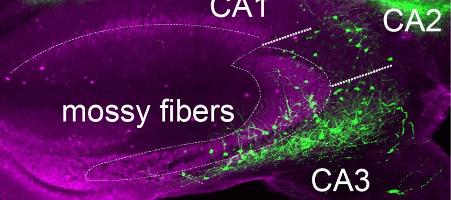
While other research teams examined the activity of interneurons in the past, most of their efforts focused on anesthetized rodents or used poorly performing techniques on moving rodents. In their recent experiments, Geiller and their colleagues set out to examine the activity of interneurons in the CA3 and CA2 regions of the mouse hippocampus as the animals were awake and moving, using a new method introduced in their previous work.
There are different types of interneurons in the brain, each characterized by distinct shapes, activity patterns and molecular signatures. In addition to exploring the possible link between interneuron activity and SWR events, the neuroscience community thus will also need to determine if some types of interneuron are more involved in this memory consolidation process than others.
“A few years ago, my colleague and I developed a method to record activity of large populations of interneurons, which has been notoriously difficult in the past, but also to molecularly identify the interneuron class that we recorded,” Geiller explained. “This is a two-step approach, based on an unconventional type of fluorescence microscopy and combined with immunohistochemistry-based labeling.”
In their experiments, Geiller and his colleagues essentially trained adult mice to perform a spatial learning task that was previously found to produce activity in the hippocampus and prompt memory consolidation. While the mice completed this task, they examined the activity of interneurons in the CA3 and CA2 hippocampal regions using their recently developed microscopy-based method.
This method allowed the researchers to characterize the activity and responses of different sub-types of interneurons as the mice were forming new memories and SWRs were generated in their brain. For instance, they found that interneurons expressing the protein cholecystokinin were less active before the SWRs took place, while interneurons expressing the protein parvalbumin were highly active after SWR oscillations.
“Recently, it has been shown that not only SWR are important for memory consolidation, but that the duration of SWR can be predictive of how well an animal remembers, or rather we should say ‘how fast an animal learns,'” Geiller said.
“Our paper suggests that the activity of cholecystokinin (CCK)-expressing interneurons (a class of interneurons) in the CA3 region of the hippocampus (where SWR are thought to originate) decreases just before SWR events, and that the magnitude of this decrease is associated with how long the SWR will be. Similarly, we found that parvalbumin (PV)-expressing basket interneurons activity is associated with the termination of SWR events, and reflective of how long the SWR was.”
The recent work by this team of researchers sheds new light on the activity of specific types of interneurons in the hippocampus before, during and after the generation of SWRs, thus contributing to the understanding of memory consolidation and learning processes. In the future, it could pave the way for further studies looking at how distinct interneuron populations influence and regulate learning related SWRs.
“In our next studies we plan to leverage the genetic specificity of these interneuron classes and to manipulate them in a temporally controlled manner with optogenetics,” Geiller added.
“We want to test whether we can artificially increase the duration of SWR events and consequently improve learning in mice. However, we know that CCK and PV interneurons are only one element of the circuit mediating SWR generation. Previous work has also implicated neuromodulation and other subcortical nuclei with this process, so it may be a long time until we fully understand the neural underpinnings of memory consolidation.”
More information:
Bert Vancura et al, Inhibitory control of sharp-wave ripple duration during learning in hippocampal recurrent networks, Nature Neuroscience (2023). DOI: 10.1038/s41593-023-01306-7
Journal information:
Nature Neuroscience
Source: Read Full Article
