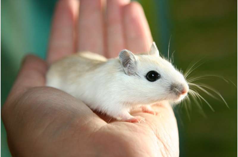
Mice are an important animal model of human vision due to the powerful genetic tools available in this species. However, mouse vision was thought to be different to that of humans because humans have a region of the retina specialized for fine details called the ‘fovea’ whereas mice do not. Researchers from the Netherlands Institute of Neuroscience (NIN) have shown that the visual cortex of mice does contain a region of enhanced visual sensitivity dubbed the ‘focea’, making the mouse a better model of human vision than previously expected. The findings were published in Nature Communications on the 29th June.
A specialization for high resolution vision
The fovea is a region in the human retina in which the light-sensitive cells are more closely packed together yielding higher resolution vision. It is used for reading and recognizing faces. Humans move their eyes three times per second to point the fovea at interesting parts of the world. Mice do not have this specialization and it was thought that they have no reason to move their eyes to ‘scan the world’ in more detail.
While studying how the retina of the mouse was mapped in cortical regions of the brain, the researchers found that the map of visual space in the cortex has a better visual resolution at a location that represents a region directly in front of and slightly above the mouse in space. They named this cortical location the ‘focea’ as it is reminiscent of the fovea of humans.
A better organized map of space
To determine the source of the better resolution the researchers measured the responses from individual brain cells using electrodes. The cells at the focea did not appear to the differ from other cells. “Initially we were puzzled” explains Matthew Self, lead researcher on the project, “we were seeing a very clear focea in the map, but nothing when we measured the data from single-neurons. We realized that the focea could originate from regions of the cortex where the maps are better organized across cells”.
To study this, the researchers measured maps of space across thousands of cells using a two-photon microscope. The results confirmed their hypothesis; maps of space in the focea were well-organized with neighboring cells responding to neighboring parts of space. In contrast, the maps outside the focea were more scattered. The results suggested that mice might have better vision in the focea than elsewhere.
The focea in natural behaviors
Source: Read Full Article
