Schizophrenia is a complex brain disorder characterized by severe social dysfunctions. A similar but attenuated format of social dysfunction has also been found in individuals with subclinical features such as social anhedonia.
Dr. Raymond Chan’s team from the Institute of Psychology of the Chinese Academy of Sciences has recently shown that both patients with schizophrenia and individuals with social anhedonia exhibited alterations in the social brain network and diminished correlation with real-world social network size characteristics. However, these findings were done with an approach that may not be able to address the complex relationship between brain functional connectivity and real-life social behavior adequately. Moreover, it is still not clearly known whether the social brain network characteristics could predict a real-world social network.
To further clarify these issues, the researchers have conducted two studies to specifically examine the association between social brain network and real-life social network size in schizophrenia patients using a hub-connected functional connectivity approach, and they verified the prediction of the identified social brain connectivity in individuals with social anhedonia.
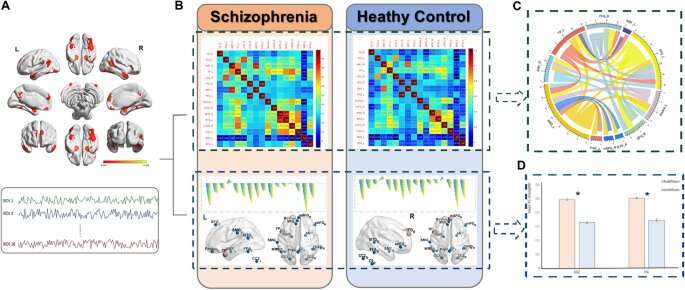
They first recruited 49 patients with schizophrenia and 27 healthy controls to undertake the resting-state brain imaging scan and complete a checklist to measure social network size. Results showed that the left temporal lobe was the only hub of social brain network, and its connected functional connectivity strength was higher than the remaining functional connectivity in patients with schizophrenia and healthy controls.
They also found that patients with schizophrenia exhibited lower association between the hub-connected functional connectivity with the real-world social network size characteristics. More importantly, they further recruited 30 patients with schizophrenia and 28 healthy controls to follow the same procedure and data analysis, and they replicated the same findings in this independent sample.
They then recruited 22 pairs of participants with high and low levels of social anhedonia. All the participants undertook the resting-state brain imaging scan and completed a checklist to measure social network size at baseline and then completed another checklist 21 months later. Results showed that social brain network characteristics could predict the change of real-world social networks in both participants with high and low levels of social anhedonia.
Notably, these results also showed a different pattern exhibited by the two groups. The topological characteristics of the social brain network predicted real-world social network change in participants with high levels of social anhedonia, whereas the functional connectivity within the social brain network predicted real-world social network change in participants with low levels of social anhedonia. Their findings also showed that the functional connectivity component centered at the right orbital inferior frontal gyrus could best predict social network change for the entire sample.
Taken together, the researchers suggest that brain regions centered at the left temporal lobe appear to be the hub region of the social brain network supporting complex social behavior. The hub-connected connectivity, compared with non-hub connected functional connectivity of the social brain network in patients with schizophrenia, affects their relationships with real-life social function.
According to the follow-up study in individuals with social anhedonia, such social brain network characteristics could predict the longitudinal change of real-world social network in individuals with high levels of social anhedonia, particularly the inferior orbital frontal gyrus functional connectivity. These findings may be important in guiding the development of non-pharmacological interventions for social function deficits in patients with schizophrenia spectrum disorders.
Source: Read Full Article