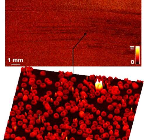
While Blu-ray discs may not have found success supplanting DVDs in the home entertainment field, Blu-ray technology is an engineering feat enabling advances in medical imaging technologies at UConn.
Guoan Zheng, the United Technologies Coorporation Associate Professor in the Department of Biomedical Engineering—a shared department with the UConn School of Dental Medicine, School of Medicine, and School of Engineering—recently tested a sensor that uses a modified Blu-ray player to produce high-quality images of biological samples. Zheng’s findings were published as the cover article in ACS Sensors last month. UConn Technology Commercialization Services has filed a provisional patent for this invention.
This technology helps address a key limitation of conventional microscopes. Microscopes provide high-resolution images at incredibly small scales. But there’s a tradeoff—in exchange for this high resolution they have a narrow field of view, meaning scientists can only see a fraction of the sample at once. The same is true in reverse: if scientists want a large field of view, they have to sacrifice the image’s resolution.
“That’s the problem with a conventional microscope—you cannot have both,” Zheng says. “And the reason comes from the design of the objective lens.”
The only way to address this issue with the current design for microscopes would be with a massive objective lens that would be prohibitively expensive, difficult to manufacture, and unwieldy to use.
Zheng’s invention combines a blood cell-coated image sensor with a modified Blu-ray drive to provide images that have both high resolution and a wide field of view.
The team smears a layer of blood cells on an image sensor. When light diffracts from an object at a large angle, the blood-cell layer redirects the light into smaller angles that the image sensor can detect. This means otherwise inaccessible high-resolution object details can be acquired using the sensor pixel array underneath the sample.
A typical Blu-ray drive consists of a rotating disc and an optical pick-up head mounted on a translation stage. The name “Blu-ray” comes from the ultraviolet 405-nanometer laser mounted on the optical pick-up head. The laser light hits the disc, and the pick-up head reads the reflected light.
In Zheng’s device, biological samples, like Petri dishes, are mounted on the rotating disc of the Blu-ray drive. The blood-coated image sensor is mounted on the translation stage. As the disc slowly rotates, the ultraviolet laser illuminates the biological samples and the resulting diffraction patterns are recorded by the blood-coated sensor for image reconstruction.
“The blood smearing process makes a thin but dense layer of blood cells on the sensor.” Zheng says. “This uniform blood-cell layer has rich spatial features for modulating both the intensity and phase of the incoming light waves, making it an ideal candidate as a computational lens.”
At the heart of the reconstruction process is a lensless coherent diffraction imaging method called rotational ptychography. By modeling the blood-cell modulation process during disc spinning, this method recovers high-resolution, large field-of-view images of biological samples with both intensity and phase information.
The recovered images allow a clinician to detect pathologies in the sample.
The device can help clinicians identify crystals in urine associated with kidney disease, for example, or parasites in blood samples.
Traditionally, in order to make a diagnosis, clinicians or lab technicians would need to stain a sample to see contrast that would reveal pathologies. This process could take hours or days. Zheng’s invention can produce a topographic height map of transparent specimens without the staining process.

“If we can recover the height or the refraction index of the sample, we might not need the staining process,” Zheng says. “Using our method, you can visualize the 3D topographic structures of transparent specimens.”
This technology has a field of view as large as a Blu-ray disc, thousands of times larger than what traditional microscopes can image, which is typically less than one millimeter in diameter. In a proof-of-concept experiment, the team monitored live bacterial cultures across an entire Petri dish and was successful in resolving individual cells.
“Compared to the limited size of a regular objective lens, our blood-cell lens can be made whatever size you want,” Zheng says. “You can, for example, smear the blood on a full-frame image sensor with a size of 36 millimeter by 24 millimeters. The imaging field of view will then be 36 millimeters by 24 millimeters.”
Another advantage of Zheng’s device is that scientists don’t need to perform focusing before capturing an image as they do with traditional microscopes. If a microscope is poorly focused, the captured image will be useless. Because Zheng’s device recovers both intensity and phase information, users can refocus the image to a specific axial plane after the data has been captured.
Right now, the device can only capture information from biological samples light can pass through. Zheng says he would like to work on developing a reflection configuration that can handle reflective samples for optical metrology applications.
Source: Read Full Article
