 insights from industryCerba Research
In this interview, Cerba Research describes how its teams were able to detect resident memory T cells in solid tumors thanks to multiplex immunofluorescence.
Can you start by explaining how T cells act as effectors cells and why this is important?
T cells are essential immune effector cells and are assigned the status of preferred targets for immunomodulation. T cells can be classified as either T effector (Teff) or T regulatory (Treg) cells.
Teff cells facilitate optimal immune responses against invading microbes and tumor antigens. Under homeostatic conditions, Tregs encourage peripheral tolerance.
However, Tregs may cause the suppression of Teff cell functions within tumors. The complex interactions between Teff and Treg determine the outcome of tumor-specific immune responses. Favorable survival in several cancer types has been associated with high Teff to Treg cell ratios as established by various methods such as flow cytometry or immunohistochemistry on serial sections.
Multiplex immunofluorescence gives a technical advantage by enabling the detection of co-expression and spatial organization of multiple targets within a preserved tissue architecture on a single slide.
What was the main objective of your study?
The main objective was to demonstrate the utility of Histalim’s HISTOPROFILE® -Treg human and mouse multiplex immunofluorescence panel to identify Treg and Teff cells in situ.
What methods were used?
Throughout the study, various methods were applied, including panel design and validation of targets and the sequential multiplex protocol with Opal® (Akoya Biosciences) fluorophores on the BOND RX® (Leica) slide stainer.
Multispectral images of subcutaneous murine Patient-Derived Xenograft (PDX) Model, human tissue microarrays, and non-small cell lung carcinoma (NSCLC) tumors were acquired with the VECTRA® PolarisTM (Akoya Biosciences) slide scanner.
Could you explain how panel validation is performed?
Panel validation is conducted using a simplex and multiplex staining comparison. Simplex slides were stained for the individual biomarkers in a simplex protocol and compared to a serial section stained with the multiplex IHC.
Staining concordance between the multiplex slide and simplex slides was determined by running the analysis with HALO® after multispectral deconvolution. The percentage of positive cells from the simplex slide was compared to the multiplex slide yielding satisfactory results for both HISTOPROFILE® -Treg panels.
What were the results yielded during the study?
After applying the Histoprofile®-Treg human panel to three non-small cell lung carcinoma (NSCLC) tissues using the Leica Bond RX, multispectral images were captured using the Vectra Polaris.
We deconvoluted the images acquired with INFORM software, allowing us to detect CD3/CD8 double-positive cells and Tregs (CD3+, CD4+, CD25+, FoxP3+) using the HALO module.
The variability of Teff/Treg can be easily appreciated between the samples.
How did the study inform your findings, and what was one of your conclusions during the study?
During the study, the approach demonstrates the effective power of Histalim’s Histoprofile® Treg multiplex immunohistochemistry and how it best serves to identify and quantify numerous immune cell populations on a single tissue. This reveals significant application potential for this method across a broad range of tissues in clinical and preclinical settings.
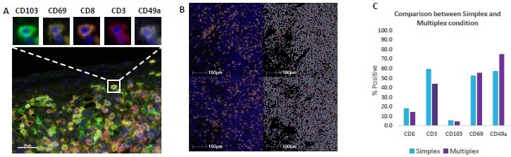
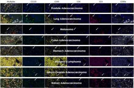
Simplex slides were stained for each individual biomarker in a simplex protocol and compared to a serial section stained with the HISTOPROFILE® TRM multiplex panel Staining concordance between the multiplex slide and simplex slides was determined by analysis with HALO® without multispectral deconvolution A) Example image of a multiplex stained healthy human tonsil The co expression of the five markers in the panel CD103 CD69 CD8 CD3 CD49 a can be appreciated in the zoom B) Example images corresponding to the multiplex slide for CD8 ( and the simplex slide ( and the corresponding HALO® masks identifying positive cells C) The percentage of positive cells from the simplex (blue bars) and multiplex panel (purple bars) slides showing comparable staining profiles between the slides. The precision of the protocol was determined by intra run repeatability analysis and inter run reproducibility analysis (data not shown). Image Credit: Cerba Research
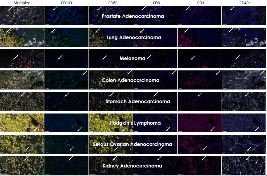
Image Credit: Cerba Research
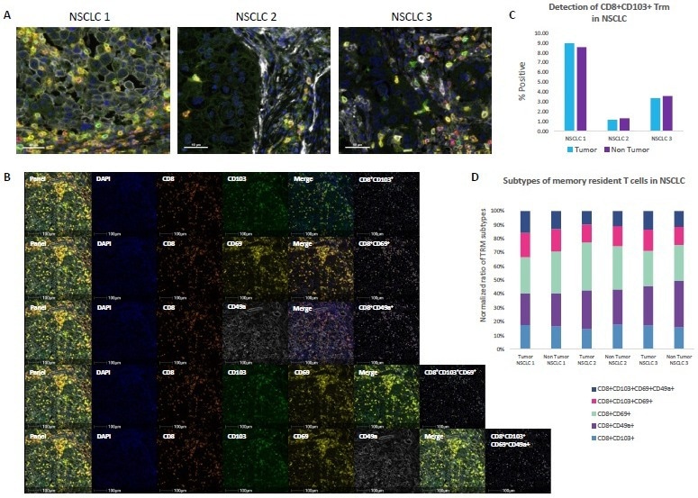
Three NSCLC samples were stained with the HISTOPROFILE® TRM panel and imaged with the VECTRA® PolarisTM Whole scan multispectral images were analyzed with HALO® A) Example of the multiplex panel on each of three NSCLC samples: CD8 (orange) CD103 (green) CD49a (white) CD69 (yellow) CD3 (red). Scale bar 40 µm In the literature, TRM have been phenotyped by different combinations of the markers used in the panel The markers were combined to analyze different TRM subtypes CD8+/CD103, CD8+/CD49a, CD8+/CD69, CD8+/CD103+/CD69, CD8+/CD103+/CD69+/CD49a in tumoral and non tumoral areas of the three NSCLC human samples B) Example images of the HISTOPROFILE® TRM panel and the individual populations with the fluorescent image and the corresponding cell masks for the different subtypes of TRM cells Scale bar 100 µm C) The frequency of memory resident T cells (CD8+ CD103+) in the tumor region (blue bars) and non tumoral area (purple bars) was analyzed D) The normalized distribution of different subpopulations of resident T cells in the tumor and non tumor regions of the three NSCLC samples CD8+/CD103+ (blue) CD8+/CD49a+(purple) CD8+/CD69+(pale green), CD8+/CD103+/CD69+(pink) CD8+/CD103+/CD69+/CD49a+(dark blue). Image Credit: Cerba Research
About Cerba Research
For over 35 years, Cerba Research has been setting the industry standard for exemplary clinical trial conduct. Today, across five continents, with a focus on precision medicine, we are changing the paradigm of the central lab’s role in complex clinical research.
From protocol inception through development and to market, our passionate experts deliver the highest quality specialized and personalized laboratory and diagnostic solutions. Partner with us for the most efficient strategy to actualize your biotech and pharmaceutical products sooner and improve the lives of patients worldwide.
Sponsored Content Policy: News-Medical.net publishes articles and related content that may be derived from sources where we have existing commercial relationships, provided such content adds value to the core editorial ethos of News-Medical.Net which is to educate and inform site visitors interested in medical research, science, medical devices and treatments.