An inherited group of integumentary disorders, Ichthyoses lead to the development of fish-scale-like skin. They are also known as disorders of keratinization (DOK) and encompass a wide range of characteristic features. These include xerosis, erythroderma, and recurrent infections.
Clinical data, analyses (such as the steroid sulfatase activity test), skin biopsies, and genetic results are used to guide the diagnostic process. However, despite significant advances in the understanding of the pathophysiology of these diseases, no cure has yet been discovered.
The treatment is multidisciplinary, and it incorporates ichthyosis patient organizations from all over the world. Moisturizing lotions are still the mainstay of treatment.
 Image Credit: Surasak muangsuk/Shutterstock
Cause
Ichthyoses involve a group of genetic disorders, caused by the inheritance of a fault or defective gene. Several mutations linked to different types of ichthyoses have been discovered over the years. Ichthyoses have been linked to mutations in more than 50 genes.
Classification
Based on pathophysiology, mode of inheritance, and clinical symptoms, the Ichthyosis Consensus Conference developed a classification consensus for DOK in 2009. Ichthyoses are divided into two groups by this nomenclature method. These are non-syndromic and syndromic forms. The disease manifests phenotypically only in the skin in non-syndromic ichthyoses, whereas syndromic forms are coupled with the involvement of other organ systems. Common ichthyoses, keratinophilic ichthyosis, autosomal recessive congenital ichthyosis (ARCI), and other non-syndromic ichthyoses are examples of non-syndromic forms. Syndromic ichthyosis, includes CHILD syndrome, Conradi-Hunermann-Happle syndrome, and Sjögren-Larsson syndrome.
Ichthyosis Vulgaris (IV)
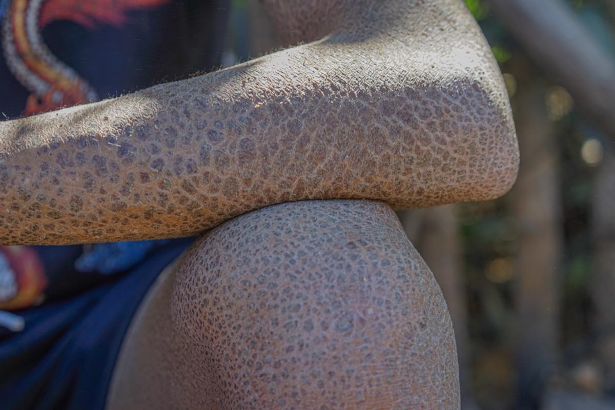
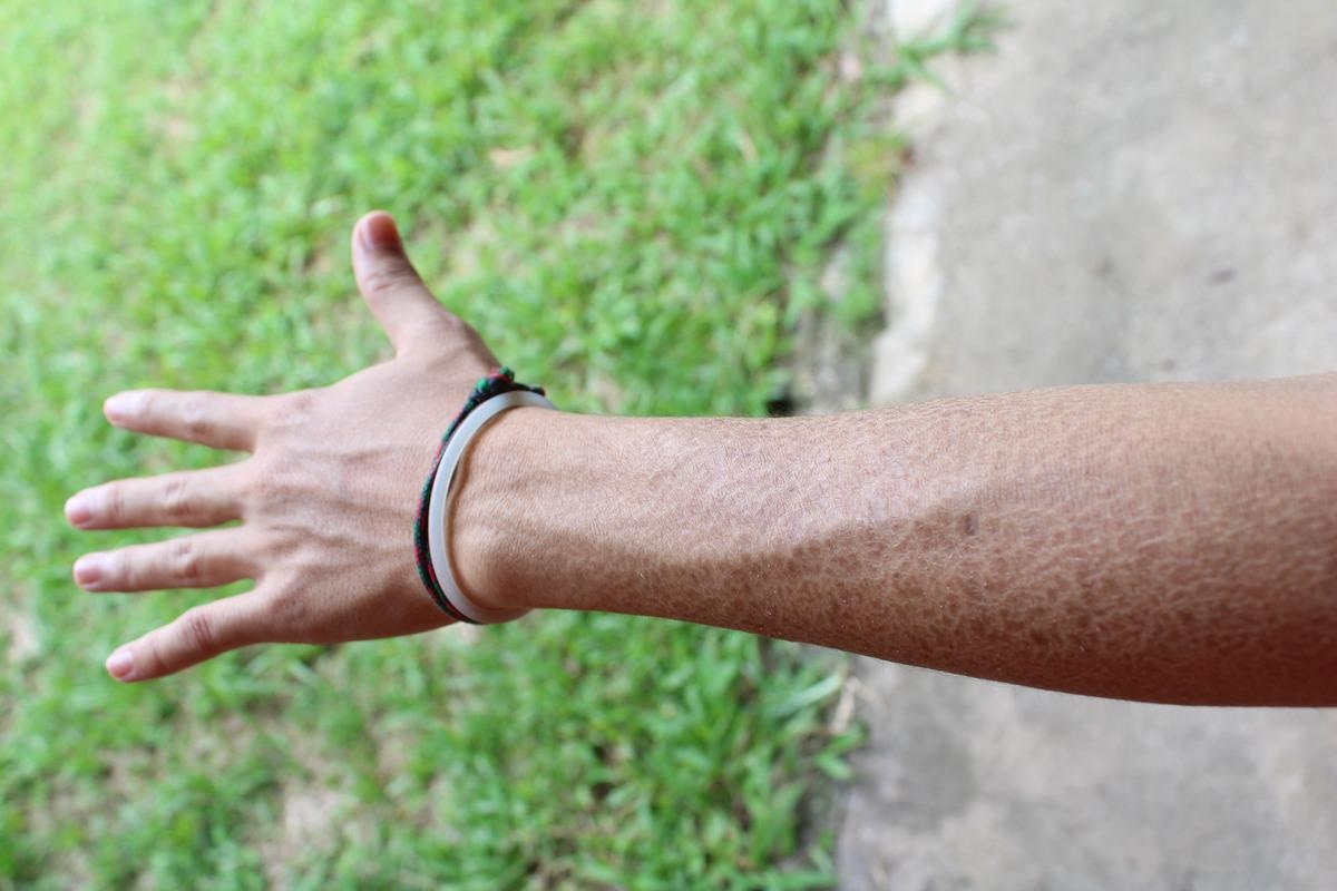
With an estimated prevalence of 1 in 250 to 1 in 1000 births, IV is the most frequent form of non-syndromic hereditary ichthyosis. It is the mildest kind of non-syndromic hereditary ichthyosis. Generalized xerosis and fine white to grayscale are common clinical findings around the age of two months. The abdomen, chest, and extensor surfaces of the extremities have the most scales. Keratosis pilaris, as well as hyper-linearity of the palms and soles, are common symptoms of IV. Autosomal dominant mutations in the filaggrin gene (FLG) cause the condition. This gene is required for epidermal differentiation and the development of the skin barrier. Atopic dermatitis, asthma, and allergies are more common in IV patients. European, African, and Asian populations have all been found to have population-specific FLG variants of IV/AD.
 Ichthyosis vulgaris. Image Credit: NUGRAHA DIPA/Shutterstock.
X-linked recessive ichthyosis (XLRI)
The second most frequent kind of inherited ichthyosis is XLRI. Males have a prevalence of 1:2000 to 1:6000. The clinical manifestations of XLRI are frequently mistaken for Ichthyosis Vulgaris. Changes in the STS gene, which encodes steroid sulfatase, cause X-linked recessive ichthyosis. Symptoms usually start as generalized desquamation and xerosis in the newborn period and proceed to fine scaling of the trunk and extremities in childhood. Patients develop a brownish, polygonal, plate-like scale that adheres to the skin tightly over time.
Autosomal Recessive Congenital Ichthyosis (ARCI)
Congenital ichthyosiform erythroderma (CIE), harlequin ichthyosis (HI), and lamellar ichthyosis (LI) are among the genetically and phenotypically variable group of diseases known as autosomal recessive congenital ichthyosis (ARCI). The incidence of ARCI is estimated to be one in every 200,000 births. Neonatal dehydration, skin infections, ectropion, eclabium, and hypohidrosis with extreme heat intolerance are common clinical signs of ARCI types. The ARCI is categorized into three major phenotypes and three minor subtypes in clinical terms. Although the Online Mendelian Inheritance in Man (OMIM) database lists 11 genetic subgroups for ARCI.
Harlequin ichthyosis (HI) is the inherited ichthyosis with the most severe phenotype. It can be fatal on rare occasions. Loss-of-function mutations in the ABCA12 gene, which genes for an ATP-binding cassette (ABC) transporter, induce harlequin. This gene is required for lipid transport into lamellar granules and is essential for cornification and the creation of lipid barriers. The ears of neonates with HI have thick, armor-like scaling with eclabium, significant ectropion, and flattening. Although some patients with HI die during the neonatal era, progress in neonatal intensive care and early treatment with systemic retinoids has been demonstrated to enhance survival.
LI is a less severe form of ichthyosis than HI. The degree of skin problems, such as hyperkeratosis and scales, differs from one patient to another. The scales that cover the majority of the body surface in LI are big, thickened, and dark grey or brown. Generally, erythroderma is not included in LI, though there have been a few reports of extremely mild erythema.
Another minor ARCI variant is bathing suit ichthyosis (BSI). In South Africa, the name "bathing suit" ichthyosis was used to describe this peculiar phenotypic of lamellar ichthyosis. It is distinguished by a distinct pattern of lesions on the trunk. The most proximal regions of the upper limbs, the scalp, and the neck are also affected, but not the central face or extremities. TGM1 deficit with heat-dependent TGM1 dysfunction can cause BSI.
Keratinopathic ichthyosis
Keratinopathic ichthyosis refers to a collection of diseases caused by mutations in the keratin gene family. Epidermolytic ichthyosis is the most common type of keratinopathic ichthyosis (EI). Surface EI (SEI), annular EI (AEI), and ichthyosis Curth-Macklin are minor forms.
EI is characterized by skin fragility and is caused by autosomal dominant mutations in the KRT1 and KRT10 genes. AEI is a rare phenotypic form of EI induced by a single KRT10 mutation. It is characterized by the production of blisters during birth. Another unusual illness is ichthyosis Curth-Macklin, which is characterized by extensive spiky or verrucous hyperkeratosis over the trunk and extensor surfaces of the extremities. Autosomal dominant mutations in KRT1 cause it.
Ichthyosis with Confetti (IWC)
Ichthyosis with confetti (IWC), also known as ichthyosis variegata, is an autosomal dominant congenital ichthyosis. IWC is classified as non-syndromic ichthyosis in the current categorization. It is characterized by global ichthyosiform erythroderma, which appears at birth. IWC is extremely rare with a prevalence of less than 1 case in 1,000,000 births.

 Read Next: Effects of Dry Skin
Diagnosis and Treatment
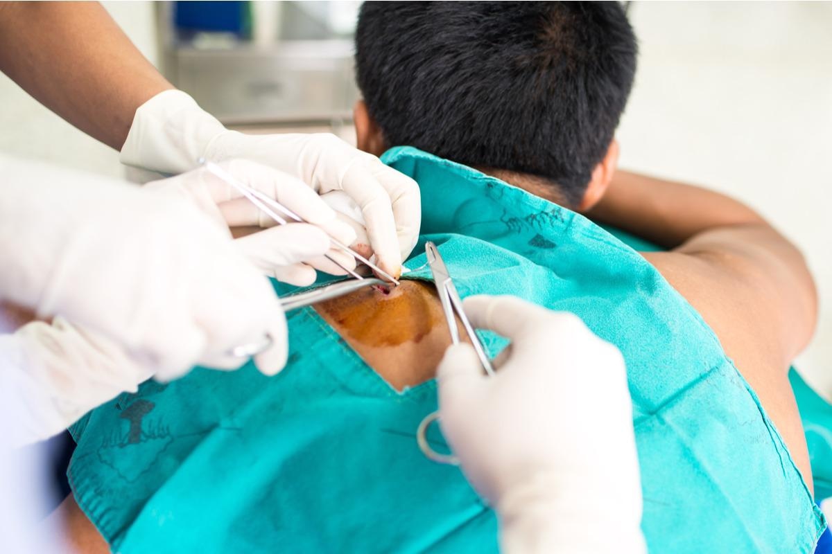
A combination of clinical data, patient history, laboratory analyses, genetic tests, and skin biopsies are used in the diagnosis.
Depending on the severity of the ichthyosis, hydration and lubrication are combined with keratolytic and keratinocyte differentiation modulators. Creams and ointments with low quantities of salt, urea, or glycerol can be used to hydrate the skin. The keratolytic properties of retinoids can help patients with LI, epidermolytic hyperkeratosis, or CIE shed their skin and avoid additional hyperproliferation. The majority of ichthyosis treatments work to increase the skin's barrier function.
Bathing and careful use of the bland creams and ointments outlined previously are important components of an ichthyosis patient's daily regimen. Bathing with water or a mild cleanser daily and applying basic emollients immediately afterward (as well as throughout the day) helps to seal in moisture.
 Skin biopsies can be used for the diagnosis of Ichthyoses. Image Credit: Chanpen Supagoson/Shutterstock
The importance of genetic counseling in the management of Ichthyoses is highly significant. Genetic counseling can serve as a source of all required information for the patient and their family. Further study into lotions, ointments, and even cosmetics have the potential to improve the quality of life for patients who are suffering from physical discomfort and social stigma as a result of the disease. Furthermore, the development of gene therapy holds enormous potential for treating patients with these illnesses and should be pursued.
References
- Oji, V., & Traupe, H. (2009). Ichthyosis. American journal of clinical dermatology, 10(6), 351-364. https://doi.org/10.2165/11311070-000000000-00000
- Guerra, L., Diociaiuti, A., El Hachem, M., Castiglia, D., & Zambruno, G. (2015). Ichthyosis with confetti: clinics, molecular genetics and management. Orphanet journal of rare diseases, 10, 115. https://doi.org/10.1186/s13023-015-0336-4
- Guerra, L., Diociaiuti, A., El Hachem, M., Castiglia, D., & Zambruno, G. (2015). Ichthyosis with confetti: clinics, molecular genetics and management. Orphanet journal of rare diseases, 10, 115. https://doi.org/10.1186/s13023-015-0336-4
- Marukian, N. V., & Choate, K. A. (2016). Recent advances in understanding ichthyosis pathogenesis. F1000Research, 5, F1000 Faculty Rev-1497. https://doi.org/10.12688/f1000research.8584.1
- Takeichi, T., & Akiyama, M. (2016). Inherited ichthyosis: Non-syndromic forms. The Journal of dermatology, 43(3), 242–251. https://doi.org/10.1111/1346-8138.13243
- Limmer, A. L., Nwannunu, C. E., Patel, R. R., Mui, U. N., & Tyring, S. K. (2020). Management of ichthyosis: a brief review. Skin Therapy Lett, 25(1), 5-7.
Further Reading
- All Rare Disease Content
- What is a Rare Disease?
- Teaching old drugs new tricks – drug repurposing for rare diseases
- What is Agnosia?
- What is Ameloblastoma?
Last Updated: May 10, 2022

Written by
Aimee Molineux
Aimee graduated from Oxford University with an undergraduate degree in Japanese and Korean Studies, with an exchange year at Kobe University in Hyogo, Japan. Throughout her studies, Aimee took part in various internships, gaining an interest in marketing and editorial work along the way.In her personal time, Aimee can be found either attempting to cook, learning how to code, doing pilates, as well as regularly updating her pet hamster’s Instagram account.
Source: Read Full Article
