Endocarditis is a life-threatening disease that involves inflammation of the endocardium, heart valve, and vascular intima. Endocarditis is caused by pathogenic bacteria – around 90% of the cases are caused by gram-positive cocci bacteria such as streptococci, staphylococci, and Enterococci.

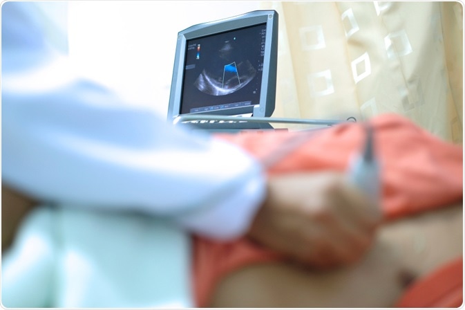
Echocardiography. Image Credit: PIJITRA PHOMKHAM/Shutterstock.com
Endocarditis – Causes, risk factors, and symptoms
Endocarditis mainly affects patients with heart disease. In patients with heart diseases, the force generated by turbulent blood flow damages the endocardium, the heart's inner lining. Next, the bacteria attach to the damaged endocardium. The tissue damage also leads to thrombus formation. The bacteria mix with the thrombus and form clumps called vegetation.
Vegetation is a specific indicator for the diagnosis of endocarditis. The vegetation can break and reach other organs such as the lungs, kidneys, spleen, and brain – this can cause several complications such as heart failure, stroke, and kidney damage.
Age is a significant risk factor. Endocarditis is typically observed in individuals above 60 years of age. The increased use of artificial heart valves, pacemakers, and various endovascular procedures also increase the probability of endocarditis. Illegal intravenous drug use and poor dental health are other risk factors.
The early symptoms of endocarditis can be nonspecific and varied. The most common symptoms are heart murmur, fever, and anemia.
Endocarditis is associated with poor prognosis and high mortality. Early detection and early treatment are necessary to avoid complications such as heart failure and organ embolism, which may manifest as cerebral embolism and splenic embolism.
Endocarditis – Diagnosis
Endocarditis is diagnosed by assessing the clinical symptoms along with echocardiography and blood culture tests.
The increasing use of antibiotics and the use of invasive techniques to diagnose heart disease have made diagnosis endocarditis a challenge. The symptoms of endocarditis have become mostly atypical, and this increases the chances of missed diagnosis.
Echocardiography in endocarditis
Echocardiography is the preferred technique for diagnosing endocarditis and is recommended to be carried out as soon as endocarditis is suspected.
How does echocardiography help detect endocarditis?
Echocardiography is an imaging technique that uses ultrasound waves to create images of structural changes in the heart.
Echocardiography helps detect vegetation and assess the range of vascular damage and the hemodynamic abnormalities in endocarditis. It also helps detect the probability of complications. Transthoracic echocardiography can rapidly and accurately detect vegetation and accurately determine the vegetation size, number, and location.
Types of echocardiography
Transthoracic echocardiography and transesophageal echocardiography are commonly used to detect endocarditis.
Transthoracic echocardiography
Transthoracic echocardiography is the recommended imaging technique for the initial diagnosis of endocarditis. The technique involves placing a transducer on the patient's chest, which delivers ultrasound waves through the chest wall to the heart.
The chances of detecting vegetation in endocarditis through transthoracic echocardiography can be reduced in conditions such as chronic obstructive pulmonary disease, structural abnormality of the chest, and obesity. Sound shadows which develop due to artificial valves can also interfere with the results. In such cases, the clinician may suspect endocarditis, but the transthoracic echocardiography may fail to detect abnormalities. Transesophageal echocardiography is recommended to dissect information in such cases.
Transesophageal echocardiography
When transthoracic echocardiography is positive or nondiagnostic, the next step is transesophageal echocardiography. In transesophageal echocardiography, the transducer is attached to a flexible tube inserted down the patient's throat and into the esophagus. Transesophageal echocardiography enables the doctor to view the heart structures more intricately. Transesophageal echocardiography is the preferred choice in complicated cases, and the presence of intracardiac device leads. Transesophageal echocardiography is highly recommended in cases with Staphylococcus aureus bacteremia, which is associated with higher morbidity and mortality.
Transesophageal echocardiography is more sensitive than transthoracic echocardiography for detecting complications. Transesophageal echocardiography has a sensitivity of 90%–100% and a specificity of 90% for the detection of vegetations. The specificity depends on the ability of the techniques to differentiate valvular vegetation from other intracardiac masses and artifacts arising in ultrasound.
Three-dimensional echocardiography
With advances in techniques, novel echocardiography techniques have emerged. Three-dimensional (3D) echocardiography is one such innovative technique.
The main challenge with two-dimensional 2D transesophageal echocardiography is selecting the maximum actual diameter of the vegetation or bacterial masses, leading to underestimation of vegetation. This challenge can be resolved with the use of 3D transesophageal echocardiography.
3D transesophageal echocardiography helps acquire infinite planes and a volumetric reconstruction of masses, enabling a better estimation of vegetation size, structure, and embolic risk.
Summary
The management of endocarditis is complex and requires a team of specialists and integrated multimodality diagnostic techniques. Echocardiography is a vital tool in the diagnosis and management of endocarditis.
Echocardiography helps decipher the structure of the heart valve and the surrounding heart anatomy and function, which, in turn, enables prompt treatment of endocarditis.
References:
- Sordelli, C., et al. (2019). Infective Endocarditis: Echocardiographic Imaging and New Imaging Modalities. Journal of cardiovascular echography, 29(4), 149–155. https://doi.org/10.4103/jcecho.jcecho_53_19
- Yuan, X. C., et al. (2019). Diagnosis of infective endocarditis using echocardiography. Medicine, 98(38), e17141. https://doi.org/10.1097/MD.0000000000017141
- Evangelista, A., et al. (2004). Echocardiography in infective endocarditis. Heart (British Cardiac Society), 90(6), 614–617.
Further Reading
- All Endocarditis Content
- What is Endocarditis?
- Infective Endocarditis
- Non-Infective Endocarditis
Last Updated: Aug 17, 2021
Source: Read Full Article
