 insights from industryRobert Tycko Acting Chief: Laboratory of Chemical PhysicsNIH/NIDDK
In this interview conducted at Pittcon 2023 in Philadelphia, Pennsylvania, we spoke to this year's recipient of the Pittsburgh Spectroscopy Award, Robert Tycko.
Could you introduce yourself and tell me a little bit about your personal background and what first attracted you to this field?
I am Rob Tycko, a research group leader at the National Institutes of Health (NIH) on the main campus in Bethesda, Maryland.
I am part of the National Institute of Diabetes and Digestive and Kidney Diseases (NIDDK). The NIH comprises around 25 institutes with somewhat different missions, most of which have basic research programs.
I am part of the basic research program at my institute, in a department called the Laboratory of Chemical Physics. It is a group of scientists with physical chemistry, biophysics, and physics backgrounds.
I primarily do nuclear magnetic resonance-based research, and I have been doing that since around 1980, when I started graduate school. What initially attracted me to the field was mathematics, as magnetic resonance has interesting conceptual and theoretical aspects based on quantum mechanics, and that is something I enjoy.
When I began my career, there was much opportunity to build new equipment because it was still in the early days of the type of nuclear magnetic resonance (NMR) I do, primarily solid-state NMR. This means NMR methods that are developed specifically for studying structural and dynamical properties and other properties of solids, as opposed to simple liquids or solutions. Traditional NMR, analytical NMR, as used by chemists, is usually liquid-state or solution NMR.
Applying NMR techniques to solids and getting molecular-level information was a relatively new field in the early 1980s and was still developing rapidly. We built a lot of our equipment, and as I enjoy working with my hands, this is how I got into the field and what attracted me to it. The other thing I like about the field, which has turned out to be true throughout my career, is that you can apply these techniques to various systems in various fields. So I have been able to contribute to various fields, including problems primarily in physical chemistry, pure physics, biophysics, and biology. Since moving to my current position at NIH about 29 years ago, I have concentrated primarily on biomolecular systems.
What is magnetic resonance spectroscopy, and what are its uses?
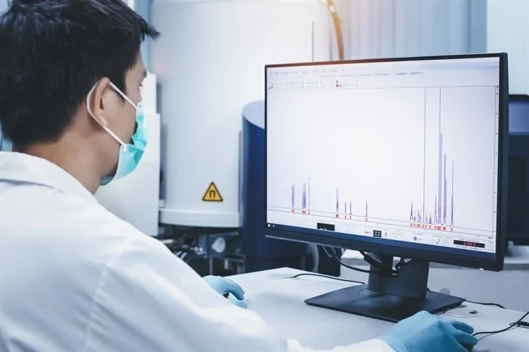
Magnetic resonance spectroscopy is a type of spectroscopy where a sample is placed in a strong magnetic field. The absorption and emission of radio wave radiation –radiation with wavelengths of centimeters to meters and frequencies of 10 megahertz up to a gigahertz–is analyzed. It is a relatively long wavelength and low energy.
Pulses of radiation are applied in modern NMR techniques, often intricate sequences of pulses that excite the motion of magnetic moments of nuclei within molecules or other materials, causing them to precess and rotate perpendicular to the external magnetic field in which the sample is sitting. We then detect the radio waves that they emit.
These signals contain lots of information about chemical structures, including three-dimensional structures and motions. Solid-state NMR measurements are also sensitive to magnetic and electronic properties and to phase transitions.
Consequently, NMR has applications in a wide variety of areas. My recent work has focused on molecular structural properties, primarily biopolymers. We have carried out much work on molecular structures of solid-like protein assemblies. These include protein tubes or filaments suspended in an aqueous solution, as non-crystalline materials.

Image Credit: S. Singha/Shutterstock.com
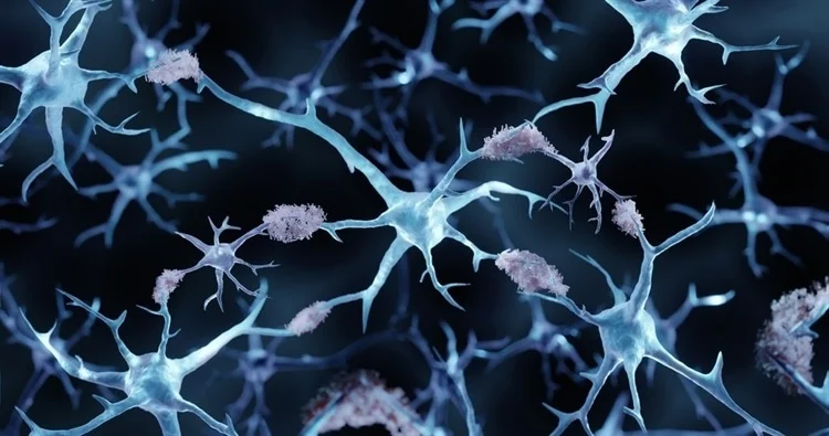
The molecular structures have biological importance, in some cases of neurodegenerative diseases, such as Alzheimer's disease. For amyloid fibrils associated with Alzheimer's disease, we could get information about the molecular structures within these non-crystalline protein assemblies that was not available from any other techniques.
The basic idea of NMR is familiar to most chemists because it is part of our training. When we study organic chemistry as an undergraduate, we learn about the fundamental principles of NMR – not the more complex techniques that have been subsequently developed. As an undergraduate studying chemistry, you are unlikely to learn much about solid-state NMR.
What is solid-state nuclear magnetic resonance?
Solid-state NMR means NMR applied to solids. In the 1950s, solids referred to solid materials interesting primarily to physicists.
In the 1960s, techniques were developed to help study molecular solids. The difference between solid and liquid NMR– the more commonly studied liquid-state NMR – is that the molecules are somehow immobilized in a solid. They do not rapidly rotate, translate, and diffuse as they are in a liquid. This affects their NMR signals, the NMR spectra that we detect in very profound ways.
In a liquid, you have very rapid tumbling and translational motions, and solvent molecules pass by the molecules you are studying. All of this makes all the molecules essentially equivalent to one another because, on average, all the molecules look the same over microseconds.
Once you immobilize the molecules, they look different from one another. The different molecules are at different orientations and could be in different environments. Those environments are very long-lived, and the differences among environments are very long-lived. These things tend to give the NMR spectra much broader resonance lines. If you do not do anything more sophisticated, the NMR spectra are very poorly resolved.
Chemists are all familiar with chemical shifts. If you have different carbon sites in an organic molecule, each has its own distinctive chemical shift when averaged over all the rotational motion. Those differences get smeared out when you superimpose an orientation dependence. Resolving the chemical shift differences among different sites within a molecule depends on special techniques.
One such technique is magic angle spinning, where you rapidly rotate your sample packed in a cylindrical rotor about a particular axis relative to the external magnetic field. That magically averages out the orientational dependence and gives you relatively sharp lines that look similar to the lines you would observe in a liquid.
This technique was initially discovered in the 1960s but has been continually improved. Samples can be spun rapidly at many kilohertz, tens of kilohertz, or even a hundred kilohertz to give you sharp lines. This is a unique technique for solid-state NMR.
There are also pulse sequences that allow you to selectively detect specific kinds of interactions that are of interest. One thing we like to do is measure distances between pairs of nuclei. The nuclei that you observe in NMR are nuclei with magnetic properties. This includes hydrogen nuclei, carbon-13 nuclei, nitrogen-15, nitrogen-14, and phosphorus-31. Certain nuclear isotopes have magnetic moments associated with them.
Depending on the distance between pairs, for a pair of nuclei, there is a coupling between the two magnets. If you take two magnets and then close them together, you can feel them interacting, repelling, or trying to twist each other. This is called a magnetic dipole-dipole coupling. We try to measure those couplings to give us information about distances.
When carrying out this magic angle spinning technique, specific pulse sequences are used to manipulate and measure the couplings.
This is something that I have worked on for many years, starting about 30 years ago with techniques that allow you to measure distances. If you measure enough distances, you can reconstruct the whole three-dimensional structure. We can now do that for complicated systems, including amyloid fibrils involved in Alzheimer's disease. That has been a big project in my lab and gives some flavor for what we can do with NMR and solid-state NMR.
How is solid-state NMR applied in biology?
Amyloid fibrils are filamentous assemblies that specific proteins and peptides form spontaneously. These amyloid assemblies are of great interest because they are associated with Alzheimer's disease, Parkinson's disease, Huntington's disease, and ALS. In the case of Alzheimer's disease, it is a particular peptide called the amyloid beta or A-beta peptide that forms amyloid fibrils in amyloid plaques within brain tissue.
These assemblies somehow contribute to the neurodegeneration that occurs in Alzheimer's disease. Exactly how that happens is something that still needs more research.

Image Credit: ART-ur/Shutterstock.com
My lab was not the first to study amyloid fibrils with solid-state NMR, but we did some initial experiments that got the field going and showed that the solid-state NMR methods that my lab and other labs had developed in the preceding five or eight years worked very well in the studies of amyloid fibrils. They provided information that was of widespread interest to the biomedical community. Many labs subsequently became involved in studies of amyloid structures, and this became one of the main applications for solid-state NMR in biology.
There are other applications, such as membrane proteins – proteins and peptides that interact with biological membranes. By interacting with the membrane, the proteins become immobilized. When studying them in an actual membrane, you cannot use traditional NMR techniques because the molecules are not rapidly tumbling and moving around. They are essentially immobilized, and you have to apply solid-state NMR techniques.
We are also working on frozen solutions. Molecules that normally behave like simple solutes in liquids are usually studied using solution NMR. In our experiments, we freeze solutions to trap intermediate structural states at various time points. Solid-state NMR is then needed to study these intermediate states. This is a new application of solid-state NMR that my lab is pioneering to study time-dependent processes of biological interest. We call it "time-resolved solid-state NMR".
We can initiate biological, biophysical, or biochemical processes in various ways, for example by mixing two solutions very rapidly or by rapidly changing the temperature of a solution. Proteins or other molecules then start to interact with one another or change their structures. We want to see what is happening with that process and how that process proceeds.
You can study these processes using other types of spectroscopy or by scattering techniques, but the information you get from NMR is uniquely sensitive to structure details. The way amino acid side chains adopt particular confirmations or interact with one another is only really shown by NMR.
We are learning new things by using time-resolved solid-state NMR. That has recently been one of my lab's main focuses. It is a new application that allows us to address biological processes that cannot be completely addressed in as much detail by other techniques.
Why is it important to push the spatial resolution limits of magnetic resonance imaging?
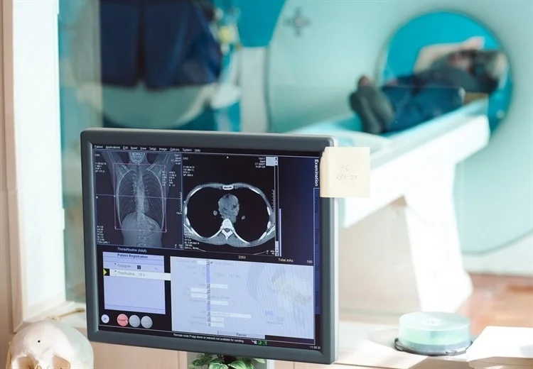
Another project in my lab that has been going on for the past 8 to 10 years is not directly related to what I have been talking about so far with magnetic resonance spectroscopy, where we are studying molecular structures. If instead you want an image of a material, assembly, or sample, there is a different set of magnetic resonance techniques, called magnetic resonance imaging or MRI. This is probably the most well-known application of magnetic resonance because many people have had MRI scans for various reasons. It was first used in the 1970s and has become an essential medical tool.
Magnetic resonance imaging is a valuable yet relatively low-resolution imaging technique. You can get images of soft tissue that you cannot get with X-rays, for example, but the resolution is not very high. It is typically a millimeter, maybe one tenth of a millimeter, which is good enough for many applications. If you need anatomical imaging, for example, to find a brain tumor or see what is happening to someone's joints or spine, the resolution is good enough to give you the information you want.
However, individual cells are much smaller, maybe 10 microns or tens of microns, and to view them, you need to get down to micron-scale resolution. For the last 40 years, researchers have attempted to push the spatial resolution further to study cells, small clusters of cells, or perhaps small tissue samples from a biopsy needle.
MRI does not currently offer a high resolution that allows you to see individual cells' arrangement or structures within cells of tiny samples. Optical or electron microscopy can be used for this instead. However, it is possible to learn something new using MRI because you see different compounds and materials and have different contrast mechanisms.

Image Credit: David Tadevosian/Shutterstock.com
Low-resolution issues in MRI come down to signal-to-noise challenges. We want to push the spatial resolution of MRI to about one micron – a thousandth of a millimeter. To do this, we need to see NMR signals from a one-micron cubed volume.
Signals from such a small amount of material are typically too weak. This is why MRI has not been commonly used to image individual cells or to examine structures inside normal cells, because of this signal-to-noise problem. You do not get enough signal when you shrink things to such small volumes.
One way to improve the signal is by using low temperatures to create stronger NMR signals. If you reduce the temperature by a factor of 10, the NMR signals increase by 10. The electronic noise also goes down, so the signal-to-noise ratio goes up. This should create higher-resolution MRI images.
At low temperatues, another phenomenon called dynamic nuclear polarization can be used to further enhance signals by another factor of a hundred.
This approach can provide massive enhancements in signal-to-noise, allowing you to get down to one micron or submicron resolution. This would make MRI more competitive with optical imaging. We are trying to develop the equipment and methods for achieving this. So far, we have been able to reach 1.7 micron resolution simple test samples. This is not very useful in real applications yet, but there is still plenty of room for further improvements.
Hopefully, in the next few years, we can get to the point where we will learn something new about individual biological cells.
What are the current challenges in NMR?
The current challenges in NMR are sensitivity and resolution. Sensitivity is an issue because we are detecting radio wave signals in NMR.
With very long wavelengths, the energies of the photons and radiation are very low compared to infrared, optical, or fluorescence spectroscopy. It is a low-energy technique with some advantages, but it means that the signals are inherently weak.
Signal enhancement is always an issue; anything you can do to increase sensitivity helps.
One thing we are doing in this MRI project and time-resolved solid-state NMR project is using low temperatures. You can freeze something in liquid nitrogen, ice, or a dry ice bath. Once you freeze it, you might as well use temperatures that are as low as possible, as you will probably not change the structure much, and the signal-to-noise will only get better. So we are pushing things to low temperatures.
The resolution of NMR is impressive compared to other types of spectroscopy. In solution NMR, people can simultaneously resolve signals from thousands of individual nuclei in a three-dimensional NMR measurement. You cannot do that with any other type of spectroscopy. Usually, you get either one signal or maybe three or four overlapping signals. In NMR, you can get thousands of signals simultaneously, each coming from individual atoms. The information content is potentially huge. For solids, the resolution is usually not as good. In solid-state NMR, because the molecules are not diffusing around or rapidly tumbling and their environments are all slightly different, the NMR lines are not as sharp. Hence, they tend to overlap with one another. The resolution is not as good. Therefore, enhancing resolution is essential.
That partly depends on technology and how fast you can spin your sample if you do magic angle spinning. It can also depend on how you prepare your sample. In the type of work I do, sample preparation methods are one area where more progress needs to be made.
One approach to improving resolution in solid-state NMR measurements on large proteins is to prepare samples in which you are only looking at a small segment at a time. This can be done by creating a protein chain by joining together several segments in which only one of them is isotopically labeled, carbon 13 or nitrogen 15 labeled. Then we can pick out the signals from the labeled segment alone. This is called segmental labeling, fragment condensation, or native chemical ligation.
On the biochemical side, if we can make this more of a routine thing, being able to make a large protein in pieces where we have isotopically labeled it so we are only looking at a relatively short segment at a time, and still be able to make the sample quantities that we need relatively quickly, that would allow us to address lots of other problems. That would allow us to address problems that are much more complicated than what we can currently do.
Are there any particular parts you are excited about that you're working on now?
I am excited about our micron-scale imaging project and the time-resolved solid-state NMR project we have worked on for the past five or six years. And we have other new projects that are also interesting, so I remain excited about our ongoing projects.
I come to meetings like Pittcon to learn about new things and to gain ideas for new projects. Then it depends on finding other people who are also excited about our projects and want to work on them in my lab.
What does it mean to you to be this year's recipient of the Pittsburgh Spectroscopy Award?
It was a great honor. I looked at the list of previous awardees, including my Ph.D. thesis advisor and his Ph.D. thesis advisor. It includes many magnetic resonance spectroscopy giants, so it has been an important award for many decades.
The most enjoyable part was inviting the other speakers, bringing them together, having dinner with them, talking to them, and listening to them talk about what they were doing.
About Robert Tycko
 Robert Tycko received his undergraduate and graduate degrees in chemistry from Princeton University and the University of California at Berkeley, respectively. After postdoctoral research at the University of Pennsylvania, he joined AT&T Bell Laboratories as a Member of Technical Staff in 1986. In 1994, he moved to the National Institutes of Health, where he is now a Senior Investigator and Acting Chief of the Laboratory of Chemical Physics, NIDDK. Tycko’s lab is best known for contributions to solid state nuclear magnetic resonance (NMR) methodology, as well as applications of solid state NMR and electron microscopy in structural studies of amyloid fibrils that are associated with Alzheimer’s disease. Recent work focuses on "time-resolved solid state NMR" techniques for studying unidirectional processes such as protein folding, peptide/protein complex formation, and amyloid self-assembly. His group is also exploring the application of low-temperature dynamic nuclear polarization in magnetic resonance imaging (MRI), recently setting a record for spatial resolution in inductively detected MRI (1.7 microns in three dimensions). Tycko is a member of the U.S. National Academy of Sciences, a fellow of the American Physical Society and the American Academy of Arts and Sciences, and a former President of the International Society of Magnetic Resonance.
 About Pittcon
Pittcon is the world’s largest annual premier conference and exposition on laboratory science. Pittcon attracts more than 16,000 attendees from industry, academia and government from over 90 countries worldwide.
Their mission is to sponsor and sustain educational and charitable activities for the advancement and benefit of scientific endeavor.
Pittcon’s target audience is not just “analytical chemists,” but all laboratory scientists — anyone who identifies, quantifies, analyzes or tests the chemical or biological properties of compounds or molecules, or who manages these laboratory scientists.
Having grown beyond its roots in analytical chemistry and spectroscopy, Pittcon has evolved into an event that now also serves a diverse constituency encompassing life sciences, pharmaceutical discovery and QA, food safety, environmental, bioterrorism and other emerging markets.
Sponsored Content Policy: News-Medical.net publishes articles and related content that may be derived from sources where we have existing commercial relationships, provided such content adds value to the core editorial ethos of News-Medical.Net which is to educate and inform site visitors interested in medical research, science, medical devices and treatments.
