The infection connection: investigating the impact on dementia risk, neuroimaging reveals
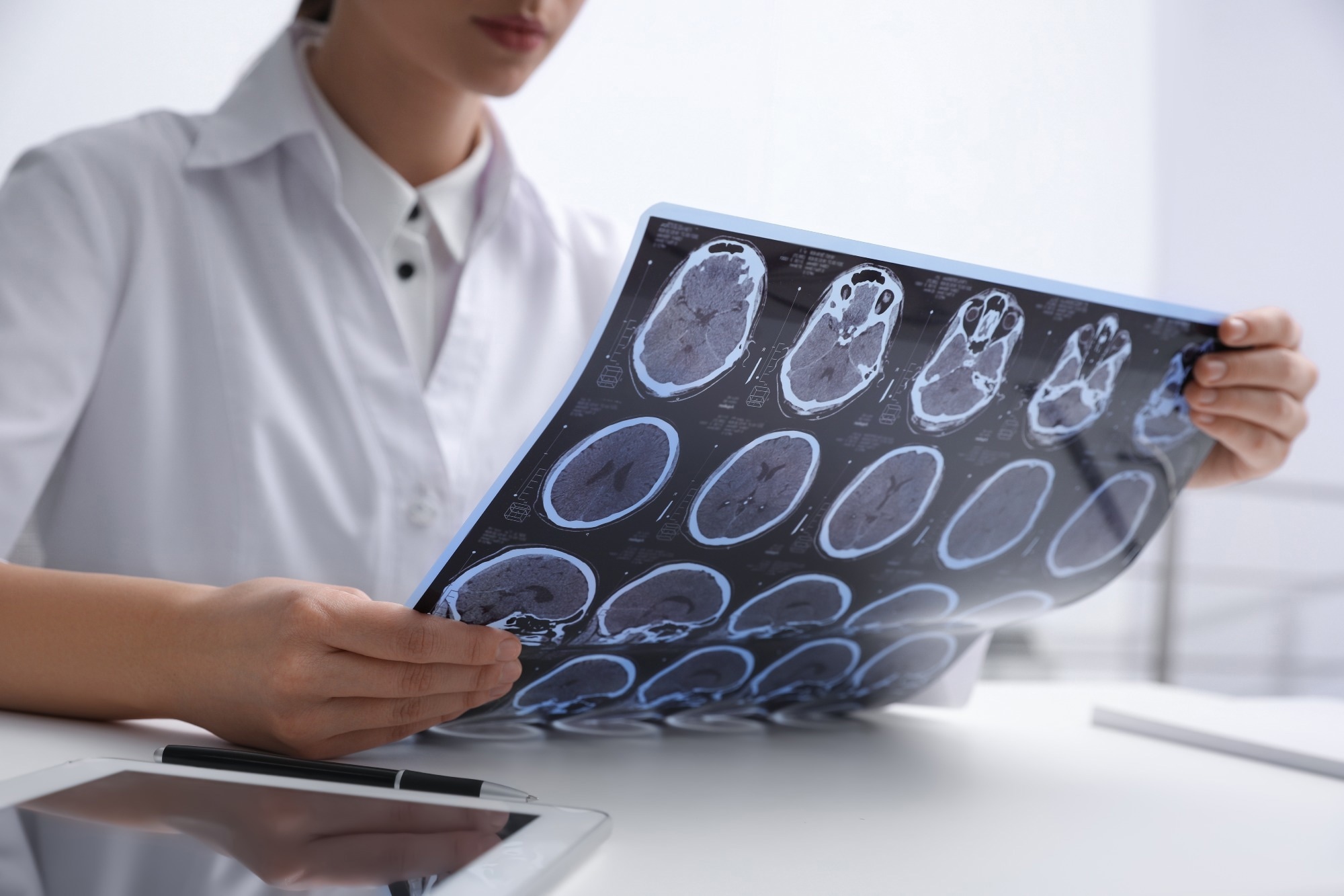
In a recent study posted to the medRxiv* preprint server, researchers compared the association between neuroimaging markers of dementia: brain volume, hippocampal volume, white matter lesions or hyperintensity volume, and antibodies to common infection across three population-based cohort studies in the United Kingdom (UK).
 Study: Common infections and neuroimaging markers of dementia in three UK cohort studies. Image Credit: NewAfrica/Shutterstock.com

 *Important notice: medRxiv publishes preliminary scientific reports that are not peer-reviewed and, therefore, should not be regarded as conclusive, guide clinical practice/health-related behavior, or treated as established information.
Background
All three markers tested in this study were associated with brain structure and pathology changes clinically relevant to subclinical dementia triggered by neurodegenerative diseases, like Alzheimer's disease (AD) and cerebral small vessel disease.
First, the researchers examined whether seropositivity and exposure to all examined pathogens were associated with neuroimaging outcomes; further, they tested these associations using the apolipoprotein E (APOE) genotype as an interaction term.
Furthermore, they assessed associations of antibody titers against each pathogen, indicating recent reactivation with these neuroimaging outcomes.
Neurotropic pathogens – such as herpes simplex virus (HSV) – can directly infect the central nervous system (CNS) cells, possibly triggering amyloid-ß pathology, neuroinflammation, and neuronal loss, all of which have been implicated in dementia etiology. However, in the absence of large-scale serology data, it remains challenging to define previous infections.
Thus, examining relationships between multiple common infections and subclinical markers of dementia prospectively – across well-characterized population-based studies – could facilitate a more comprehensive understanding of their role in dementia risk.
In addition, assessing whether pathogen exposure may interact with established risk or protective factors of dementia could provide insights into possible at-risk groups.
About the study
In the present study, researchers applied a validated fluorescence bead-based multiplex serology panel to the UK Biobank (UKB), the MRC National Survey of Health and Development (NSHD), and Southall and Brent Revisited (SABRE) to measure serum immunoglobulin G (IgG) against antigens from a wide range of pathogens with outcomes among several settings in parallel.
An adaptation of the same panel assayed 18 pathogens in 1,813 and 1,423 NSHD and SABRE participants, respectively. In UKB, they assayed 21 pathogens among 9,429 participants at baseline and an additional 260 at follow-up.
They quantified antibody responses or seroreactivity indicating prior infection for each pathogen using median fluorescence intensity units and between one and six antigens per pathogen.
To measure total exposure to multiple pathogens, they derived two pathogen burden index (PBI) scores, total PBI and neurotropic PBI indicating the sum of serostatuses to 17 pathogens and 11 neurotropic pathogens, including herpes simplex viruses (HSV), Toxoplasma gondii, and John Cunningham (JC) virus, respectively. The latter held more clinical relevance to neurological outcomes.
Next, the team derived tertiles in the serology and neuroimaging samples to group antibody responses of seropositive samples in seroreactivity analyses. Seroreactivity values formed a variety of non-normal distributions.
For NSHD and UKB, the team used neuroimaging measures assessed via brain magnetic resonance imaging five to 11 years and one to 13 after blood sampling for serology assaying, respectively. However, for SABRE, they used neuroimaging measures collected at the same time as blood sampling for serology assaying.
For all three cohorts, they quantified brain volume and hippocampal volume using in-house using Geodesic Information Flows. Likewise, they used an automatic algorithm, Bayesian Model Selection (BaMoS), for deriving white matter hyperintensities.
Further, the researchers used directly genotyped data for APOE genotypes using rs7412 and rs429358 single nucleotide polymorphisms (SNPs). Subsequently, they developed APOE e4 and APOE e2 non-carrier/carrier, where the latter were heterozygous\homozygous for the alleles e4 and e2, respectively.
Then, they used random-effects models with a maximum likelihood estimator for meta-analysis of findings across studies. Multiplying regression coefficients deduced in white matter lesion volumes analyses by 100 transformed them to sympercents.
They used the Benjamini-Hochberg procedure with an alpha of 0.05 to correct findings from meta-analyses of serostatus outcome, seroreactivity, and APOE interaction analyses and fetch the false discovery rate (FDR). An I2 statistics >50% or Q-p value<0.05 indicated significant heterogeneity in all included studies.
The team applied three linear regression models in the primary analyses to evaluate associations between serology variables and neuroimaging outcomes. While Model one included total intracranial volume and other technical covariates, Model two adjusted for Model one and age, gender, and ethnicity covariates.
Model three adjusted for model one and two covariates and social, behavioral, and lifestyle confounders. These measures reflected trackable, long-term characteristic differences.
In statistical modeling of seroreactivity analyses, they modeled tertiles as an ordinal variable and investigated pathogens with a seroprevalence >5% in all studies. The team used the same models accounting for ten genetic principal components during APOE interaction analyses to assess whether pathogen burden relationships with outcomes varied by APOE genotype, including APOE e4 and APOE e2 carrier statuses as interaction terms.
Notably, they conducted APOE e4 and APOE e2 analyses separately. The team also conducted several secondary and sensitivity analyses.
Results
The present study had 2,632 participants with available serology measures and data on at least one neuroimaging outcome, of which 438, 1,259, and 935 belonged to the NSHD, SABRE, and UKB cohorts, respectively. Likewise, the study encompassed 17 pathogens with relevant serology data.
For the APOE genotype interactions study, the authors had genetic data of 1,810 participants after quality control, of which data of 413, 593, and 804 participants came from the NSH, SABRE, and UKB cohorts, respectively. Of 593 SABRE participants, 314 and 279 were of European and South Asian ethnicities, respectively.
The authors found minimal or no evidence of any associations in most instances. Accordingly, the findings for HSV concerning neuroimaging outcomes were null though it is the most studied pathogen with an alleged link with AD.
Likewise, the findings for many other pathogens with serological markers on the study panel with alleged links to AD and other causes of dementia were null.
These findings, at least for HSV, agreed with several previous studies. Moreover, the authors found no convincing evidence of associations of pathogen burden scores derived from counts of serostatus values with neuroimaging outcomes.
However, they hypothesized that the combined burden of many pathogens drove the development of neuropathology.
In the UKB study, researchers found an association of HSV1 serostatus with incident dementia. However, the study sample of 84 incident dementia cases likely lacked the power to detect any such clinically meaningful associations.
Furthermore, the authors noted several suggestive associations. For instance, the difference of -0.07ml in hippocampal volume between VZV seropositive and seronegative individuals equated to nearly two-thirds of the lifelong-averaged effect of APOE ε4 carriage on hippocampal volume (-0.11ml).
Intriguingly, these suggestive findings were unanticipated based on the hypothesis that pathogen exposures would worsen neuroimaging metrics. Possibly, suggestive findings arose due to chance, e.g., from residual confounding.
Thus, researchers emphasized replicating these results using serology and neuroimaging data from other cohorts.
Importantly, environmental and genetic factors might be additionally affecting the observed associations. For instance, a study observed increased AD risk with higher HSV1 seroreactivity in APOE e4 carriers, and another found that cytomegala virus and Helicobacter pylori serostatuses were associated differently with whole brain volume in APOE e4 carriers.
Conclusions
According to the authors, associations of common infections with subclinical neuroimaging outcomes have barely been investigated. Thus, they did not have much data to draw comparisons or precedence. Yet, there are many ways to build upon the evidence presented in this study.
Studies with neuroimaging or clinical follow-up data that generate equivalent serology data for common infections could help estimate associations with higher precision to reaffirm (or refute) at least some of this study's suggestive findings.
Given many of these common infections are treatable and preventable by vaccination, this data could help inform strategies for improvising vaccination programs.
Furthermore, expanding the study data to include longitudinal outcomes, such as changes in white matter lesion burden and atrophy rates, could provide deeper insights into associations of common infections with neurodegeneration and pathology.
More importantly, incorporating other neuropathology measures, e.g., cerebral amyloidosis, and fluid-based neurodegeneration biomarkers, could help delineate the effects of pathogens on pathways accentuating dementia risk.

 *Important notice: medRxiv publishes preliminary scientific reports that are not peer-reviewed and, therefore, should not be regarded as conclusive, guide clinical practice/health-related behavior, or treated as established information.
- Preliminary scientific report.
Green, R. et al. (2023) "Common infections and neuroimaging markers of dementia in three UK cohort studies". medRxiv. doi: 10.1101/2023.07.12.23292538. https://www.medrxiv.org/content/10.1101/2023.07.12.23292538v1
Posted in: Medical Science News | Medical Research News | Medical Condition News | Disease/Infection News | Healthcare News
Tags: Alzheimer's Disease, Amyloidosis, Antibodies, Antibody, Apolipoprotein, Blood, Brain, Central Nervous System, Dementia, Fluorescence, Genetic, Helicobacter pylori, Herpes, Herpes Simplex, Herpes Simplex Virus, Imaging, Immunoglobulin, Magnetic Resonance Imaging, Nervous System, Neurodegeneration, Neurodegenerative Diseases, Neuroimaging, Nucleotide, Pathogen, Pathology, Serology, Single Nucleotide Polymorphisms, UK Biobank, Virus

Written by
Neha Mathur
Neha is a digital marketing professional based in Gurugram, India. She has a Master’s degree from the University of Rajasthan with a specialization in Biotechnology in 2008. She has experience in pre-clinical research as part of her research project in The Department of Toxicology at the prestigious Central Drug Research Institute (CDRI), Lucknow, India. She also holds a certification in C++ programming.