In a recent study published in Nature Cardiovascular Research, researchers assessed T-cell tolerance checkpoints observed in atherosclerosis.

Background
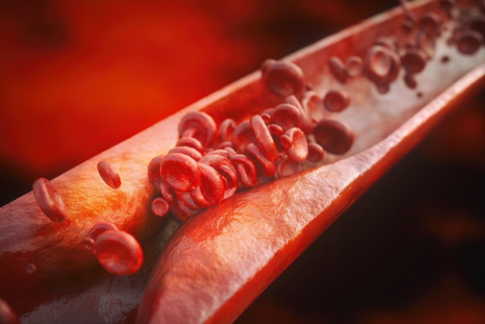
Atherosclerosis is a chronic inflammatory disease of the arteries. It is characterized by the presence of atherosclerotic plaque present in the inner layer of arteries. Plaques that rupture result in strokes or heart attacks. Significant innate immune cells that contribute to atherosclerosis have been identified.
In addition, certain subtypes of T cells, such as CD4+ regulatory T (Treg) cells and CD8+ T cells, promote or suppress the illness in mice. Yet, fundamental problems regarding T cell immunity noted in atherosclerosis are unanswered. In particular, it is unknown whether T cell responses linked with atherosclerosis are incident in the circulation.
About the study
In the present study, researchers investigated tolerance checkpoints detected in atherosclerotic plaques, lymph nodes, and tertiary lymphoid organs in mice with advanced atherosclerosis.
To investigate peripheral T cell tolerance checkpoints among mice with atherosclerosis, the team used aged Apoe -/- mice to evaluate atherosclerotic plaques, adventitial artery tertiary lymphoid organs (ATLOs), aorta-draining renal lymph nodes (RLNs), and the circulation. As controls, RLN T cells and blood of wild-type (WT) mice were sequenced using paired 5′ single-cell ribonucleic acid-sequencing (scRNA-seq). A T cell antigen receptor (TCR) and transcriptome repertoire atlas of 13,800 T cells was created.
TCR and transcriptome repertoire atlas of 13,800 T cells was created. The team examined five critical peripheral tolerance checkpoints associated with T cell immunity, such as persistence of T cell quiescence, pathways to sustain effector or memory T cell functions, Treg cell-mediated immunosuppression, antigen presentation to T cells, and tissue T cell homeostasis regulation.
Furthermore, the researchers analyzed the expression of S1pr1 along with activation- and cytokine-related genes (S100a6 and Ccl5) in naive CD8+ T lymphocytes in the blood.
Results
The study results noted 10 T cell subsets along with eight myeloid cell subsets based on the characteristic gene expression profiles of WT and Apoe-/- mice. Fluorescence-activated cell sorting (FACS) investigations of myeloid cells and T cells in the circulation of Apoe -/- and WT mice revealed comparable amounts of myeloid cell or T cell subsets. Also, blood T cells, B cells, myeloid cells, granulocyte, and natural killer (NK) cell subsets did not differ significantly between WT and Apoe-/- animals.
Omics eBook

In t-distributed stochastic neighbor embedding (t-SNE) plots, 10 T-cell subgroups emerged with respect to their differentially expressed genes (DEGs) and their primary T-cell markers. Furthermore, plaques contained large subsets of T cells and myeloid cells. In contrast to ATLOS, WT RLNs, and Apoe-/- RLNs, plaque T cell subsets had notable aberrant characteristics. Plaques comprised fewer Treg cells and naive CD4/CD8+ T cells and a higher proportion of CD8+ Tem (T cell effector memory), CD8+ Tcm (T cell central memory), and Ɣδ-T cells.
The team also validated the rise of CD4+ Tem and CD8+ Tem cells present in ATLOs compared to Apoe-/- secondary lymphoid organs (SLOs) using flow cytometry. Notably, plaques displayed a lower proportion of naive CD4+ /CD8+ T cells and a higher proportion of CD8+ Tem cells than SLOs. Therefore, a distinct disease-related and tissue-specific T-cell composition was observed.
During the transition of naive T lymphocytes to memory and effector T cells, naive T cells shifted from quiescence to an activated or memory-like state by downregulation of inhibitory checkpoint genes, such as the V-set immunoregulatory receptor (Vsir) and upregulation of activation-related transcripts. Notably, Vsir encodes a prototypical inhibitory checkpoint regulator that inhibits CD4+ T cell activation.
Hence, the downregulation of Vsir could indicate an early sign of tolerance breakdown in the naive mature T cell repertoire of the circulation. Nonetheless, total CD4+ naive T cell pool levels persisted in the circulation in elderly Apoe-/- mice compared to WT controls.
Blood nave CD8+ T cells exhibited greater S100a6 and Ccl5 levels in Apoe-/- mice than in WT mice, indicating the presence of partially activated nave CD8+ T cells in the circulation of Apoe-/- mice. Plaque-naive CD8+ T cells had elevated messenger RNA levels of Ccl5 and S100a6. Notably, there was no discernible change in the expression of these transcripts across ATLO RLNs, Apoe-/- RLNs, and WT RLNs.
Plaque-infiltrating T cells displayed 10 DEGs, comprising seven upregulated genes along with three downregulated genes in comparison to T cell clones found in RLNs and ATLOs. These ten genes constitute the plaque-inducible gene signature. The team also noted that plaque-inducible gene signatures were not affected by specific TCR sequences, as demonstrated by a comparison of three different T cell clones. These findings suggest that plaques influence T cell immunity by regulating genes associated with activation, energy consumption, and tolerance checkpoint.
Overall, the study findings provided a thorough understanding of the probable pathways behind peripheral tolerance dysfunction in late-stage atherosclerosis, thereby providing a solid framework for characterizing atherosclerosis as an autoimmune T-cell-driven disease linked with tolerance dysfunction.
Several options for future therapeutic intervention, such as restoring tolerance checkpoints and T-cell therapies, may emerge as the current studies increase understanding of an overlooked part of the pathophysiology of atherosclerosis.
- Wang, Z. et al. (2023) "Pairing of single-cell RNA analysis and T cell antigen receptor profiling indicates breakdown of T cell tolerance checkpoints in atherosclerosis", Nature Cardiovascular Research. doi: 10.1038/s44161-023-00218-w. https://www.nature.com/articles/s44161-023-00218-w
Posted in: Medical Science News | Medical Research News | Disease/Infection News
Tags: Antigen, Aorta, Atherosclerosis, Autoimmune Disease, Blood, CCL5, CD4, Cell, Cell Sorting, Chronic, Cytokine, Cytometry, Flow Cytometry, Fluorescence, Gene, Gene Expression, Genes, Heart, immunity, Immunosuppression, Inflammatory Disease, Lymph Nodes, Pathophysiology, Receptor, Research, Ribonucleic Acid, RNA, T-Cell

Written by
Bhavana Kunkalikar
Bhavana Kunkalikar is a medical writer based in Goa, India. Her academic background is in Pharmaceutical sciences and she holds a Bachelor's degree in Pharmacy. Her educational background allowed her to foster an interest in anatomical and physiological sciences. Her college project work based on ‘The manifestations and causes of sickle cell anemia’ formed the stepping stone to a life-long fascination with human pathophysiology.
Source: Read Full Article
