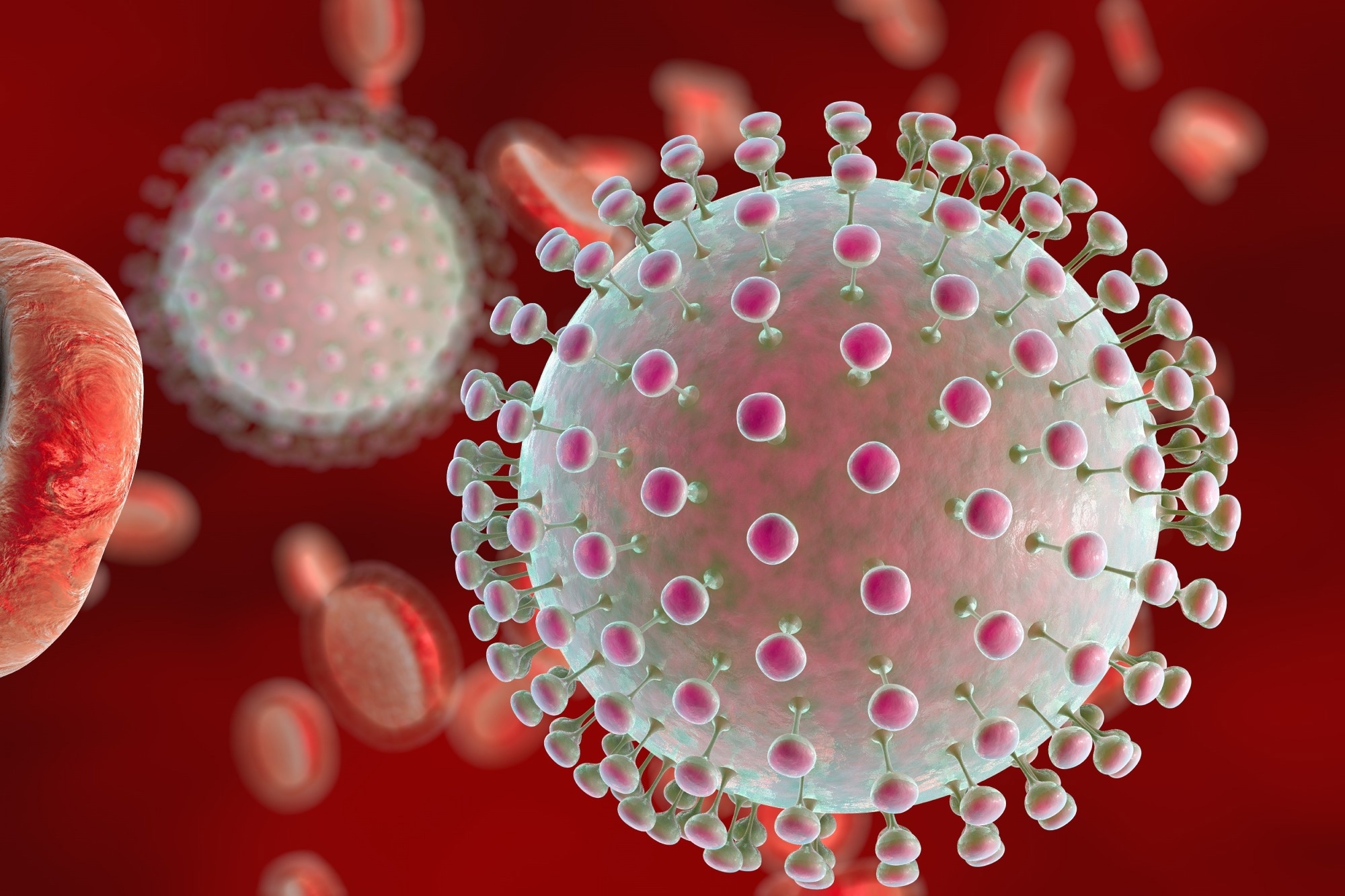
In a recent study posted to the bioRxiv* server, researchers designed two dumbbell-1 (DB-1) mutant Zika virus (ZIKV) infectious clones termed ZIKV-TL.PK and ZIKV-p.2.5’. While the former disrupted DB-1 tertiary folding, the latter altered the formation of DB-1 secondary structure. They used these mutants to investigate the contribution of the DB-1 structure to ZIKV pathogenesis.

Background
Immunology eBook

Native to Uganda, ZIKV, a virus of the Flavivirus genus, caused an outbreak in Brazil during 2015-2016, where it caused “congenital Zika syndrome” after transmitting vertically from mother to fetus. Rapidly changing climate and human distribution could expand its outbreaks into regions beyond the equator, posing a risk to human health worldwide. Also, currently, there are no antivirals or vaccines for ZIKV.
ZIKV contains multiple conserved ribonucleic acid (RNA) structures in its 3’ untranslated region (UTR), including DB-1. Previous studies have demonstrated that the DB-1 structure is crucial for its replication and cytopathic effect (CPE). However, studies have not yet uncovered the structural-functional role of DBs and the mechanism they use to enhance ZIKA pathogenesis.
About the study
In the present study, researchers thoroughly examined how the DB-1 nested in 3’UTR of ZIKV contributed to its pathogenesis. They used the three-dimensional DB structure from the Donggang virus to introduce targeted mutations and disrupt the secondary and tertiary structure of DB-1 in ZIKV. It eliminated the native role of this structure while maintaining the full-length sequence of the 3’UTR. The study investigations worked on the hypothesis that the ZIKV DB-1 structure regulated its genomic replication.
Further, the team investigated sub-genomic flaviviral RNA (sfRNA) formation by both DB-1 mutants in cell culture using mammalian A549 cells. To this end, they first infected these cells with ZIKV mutants at a multiplicity of infection (MOI) of 0.25. Next, they harvested cellular RNA at three-time points, 12, 24, and 48 hours post-infection (hpi). Finally, northern blot analysis with a ZIKV DB-1-specific probe helped assay sfRNA formation.
Note that sfRNAs play a critical role in viral pathogenesis, and their absence could result in reduced CPE, subsequently, ZIKV pathogenesis. Furthermore, they assayed ZIKV DB mutant-infected A549 cells for cell viability and caspase activation. Finally, the researchers used a mouse model to determine the attenuation of both DB-1 mutant clones following infection.
Study findings
Analyzing the viral kinetics of ZIKV mutants in cell culture revealed that their genomic replication was comparable to the ZIKV-wild type (WT) virus. However, they exhibited much reduced infectious virion production or ZIKV CPE resulting in attenuation, both in vitro and in vivo. More importantly, densitometry analysis revealed that both ZIKV mutants had decreased levels of all sfRNA species compared to ZIKV-WT. Irrespective of genome replication, mutation(s) in the 3D RNA structure of DB-1 resulted in a marked decrease in sfRNA levels during infection.
On the other hand, the researchers noted a significant surge in cell viability of both the mutants compared to ZIKV-WT due to weak caspase-3 activation. Further, they showed that type I interferon (IFN) treatment substantially limited replication of the ZIKV-P.2.5’ mutant without altering interferon-stimulated gene (ISG) expression.
In a mice model, ZIKV-DB-1 mutants exhibited reduced morbidity and mortality compared to the ZIKV-WT virus due to weakened genomic replication in the brain tissue. Future studies should examine tissue-specific attenuation induced by ZIKV mutants to inform vaccine development for flaviviruses.
Conclusions
To summarize, the study demonstrated the significance of the flavivirus DB-1 RNA structure in maintaining sfRNA levels during ZIKV infection. The study also showed, using a mouse model, how sfRNA facilitates Caspase-3-dependent ZIKV-induced CPE and pathogenesis.
*Important notice
bioRxiv publishes preliminary scientific reports that are not peer-reviewed and, therefore, should not be regarded as conclusive, guide clinical practice/health-related behavior, or treated as established information.
- Monica E. Graham, Camille Merrick, Benjamin M. Akiyama, Matthew Szucs, Sarah Leach, Jeffery S. Kieft, J. David Beckham. (2023). Zika virus dumbbell-1 structure is critical for sfRNA presence and cytopathic effect during infection. bioRxiv. doi: https://doi.org/10.1101/2023.01.23.525127 https://www.biorxiv.org/content/10.1101/2023.01.23.525127v1
Posted in: Medical Science News | Medical Research News | Disease/Infection News
Tags: Assay, Brain, Cell, Cell Culture, Gene, Genome, Genomic, in vitro, in vivo, Interferon, Mortality, Mouse Model, Mutation, Ribonucleic Acid, RNA, Syndrome, Vaccine, Virus, Zika Virus

Written by
Neha Mathur
Neha is a digital marketing professional based in Gurugram, India. She has a Master’s degree from the University of Rajasthan with a specialization in Biotechnology in 2008. She has experience in pre-clinical research as part of her research project in The Department of Toxicology at the prestigious Central Drug Research Institute (CDRI), Lucknow, India. She also holds a certification in C++ programming.
Source: Read Full Article
