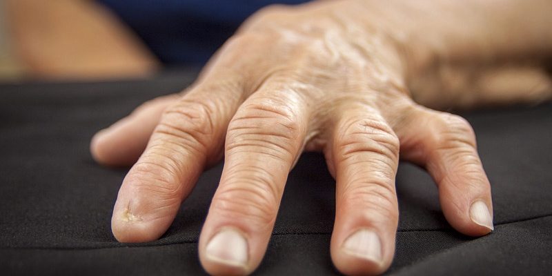
LEIPZIG, GERMANY — With age comes illness: Cancer, cardiovascular and neurodegenerative diseases, increased infections, and autoimmune diseases such as rheumatism become more common. This is because the immune system also ages. In the case of autoimmune diseases, this aging happens particularly quickly.
“There is this phenomenon of premature aging of the immune system,” said Cornelia Weyand, PhD, director of the Center for Translational Medicine at Stanford University, Stanford, California, at the German Rheumatology Congress 2023 in Leipzig, Germany. In healthy people, the immune system begins to age at age 20. From that point on, the thymus gland, which reaches peak function at 14-15 years old, plays an increasingly minor role. “At age 50 years, the aging process of the immune system gains momentum.”
“What’s good about this is that the T- and B-cells age together, but all a little differently, each system by itself,” said Thomas Dörner, MD, PhD, head of consultation hours for clinical hemostaseology at the Charité University Hospitals in Berlin, Germany.
While the reduced formation of naïve T-cells can be attributed to the regression of the thymus gland, the naïve B-cells are a consequence of age-related, fatty bone marrow degeneration. The influence of adipocyte-derived tumor necrosis factor (TNF)-α also causes the bone marrow to develop B-cells more and more weakly and slowly. “Through this [process], the preimmune range of B-cells decreases and becomes less healthy than in a young person.”
‘Inflamm-aging’
“In the periphery, we have identified a process we call inflamm-aging, where the cytokines interferon-γ, interleukin (IL)-10, and IL-17 play a predominant role. This also alters the primary and secondary immune response,” said Dörner. Here, decreasing stimulation via the B-cell receptor by aging T-lymphocytes makes a difference.
As we age, the immune system restructures itself completely. “Protective immunity regresses and the inferior immunity emerges,” explained Weyand. Wounds heal more poorly, the protective action against infections and above all malignancies, as well as the immune response to vaccinations, decreases.
The increased occurrence of neurodegenerative, cardiovascular, and autoimmune diseases is not due to a loss of function, but rather to newly gained, undesired functions. These are associated with inflammatory changes. Hence, the term “inflamm-aging.”
With the B-cells, functional germinal centers in the lymphoid organs and protective antibodies become rarer, and age-associated B-cells accumulate. As Dörner emphasized, these cells are not under the command of the B-cell receptor and are independent of the cytokine BAFF (B-cell activating factor). Instead, they react to signals that are sent from the toll-like receptors 7 and 9.
This potentially also explains the increased development of autoantibodies in older people and the association of viral and autoimmune diseases. This means that age-associated B-cells develop more frequently, such as with rheumatoid arthritis (RA), scleroderma, and systemic lupus erythematosus. “There are good data that show that they are triggered by infections and that they are specialized to form autoantibodies,” Weyand also said about the age-associated B-cells.
‘Bad Old T-cells’
Under the influence of genetic stop-and-go signals, the composition of the T-cell population also changes over the course of our lives. It becomes less diverse. T-helper cells become less common, and as a result, terminally differentiated effector memory T-cells become more common. According to Weyand, herein lies the problem. “These cells are not just lazy, old cells that sit around. Unfortunately, they are also malicious. What we see in both the T- and B-cell systems is that they become increasingly innate with age,” he said. “They are not quite so precise or good.”
In turn, myeloid cells are less active in old age due to phagocytosis and antigen presentation, and they get more mutations. They are released more often from the bone marrow, produce more cytokines, and essentially contribute to inflamm-aging.
Power Sources Fail
In her cellular and microbiological investigations, Weyand has devoted a lot of time to studying why T-cells age prematurely in patients with RA. The key was in the cellular microbiology. “We learned how the T-cell aging process translates into metabolic reprogramming of the T-cells — how a good, strong, and protective T-cell transforms into a disease-inducing T-cell.”
At the center of premature T-cell aging in RA are disrupted mitochondrial function and insufficient communication of the mitochondria with the lysosomes and the endoplasmic reticulum.
T-cells of RA patients (RA T-cells) contain less MRE11A, compared with those in healthy people. This is a nuclease that allows the repair of breaks in DNA. If MRE11A is inhibited, then senescent T-cells accumulate and form proinflammatory cytokines such as IL-B, IL-6, and TNF. “This is the trio that we rheumatologists are always concerned with.”
Since mitochondrial DNA repair is essential for maintaining mitochondrial fitness, the cellular power sources in patients with RA cannot provide as much energy in the form of adenosine triphosphate as in healthy people. “Metabolically, they are not so fit,” Weyand said.
Inflammatory Cell Death
In fact, all metabolic pathways in the T-cells are reduced. The bioenergetic failure has consequences. “Unfortunately, as the mitochondrion ages, its DNA leaks into the cytosol,” explained Weyand. “Cells do not like this.” This is because DNA activates inflammasomes in the cytosol via caspase-1. This process results in a highly inflammatory cell death: pyroptosis. Subsequently, there is no trace of the cells in the tissue. “RA patients’ synovial tissue is a graveyard of dying T-cells.”
In the lysosomes, the cells’ “intestine,” problems arise because patients with RA can no longer activate the adenosine monophosphate-activated protein kinase enzyme. It does not receive the lipid tail it needs to take its position as energy sensor on the lysosomal membrane. As a result, its antagonist, mTOR — both usually keep each other in check — gains the upper hand. According to Weyand, “mTOR has a party.” It activates and stresses the cells.
Additional changes affect the endoplasmic reticulum (ER). “This is where all of your proteins are synthesized and packaged to migrate from within to outside the cell, or to the cell membrane.” Compared with healthy T-cells, RA T-cells have around 50% more ER. “The less that mitochondria work, the larger the ER. It gets really fat.”
Mitochondria communicate with the ER via aspartate, oxaloacetate, and malate. In so doing, they control their size. RA T-cells appear to be aspartate deficient. In animal models, amino acids had an anti-inflammatory effect.
When sequencing the mRNA bound to the ER, Weyand and her colleagues encountered the building blocks for TNF. “There is more than three times as much mRNA as TNF. It transforms these T-cells into TNF superproducers,” said the rheumatologist. “No wonder this kind of cell is proinflammatory — it forms precisely that cytokine on which you focus every day.”
This article was translated from Medscape’s German edition.
Source: Read Full Article