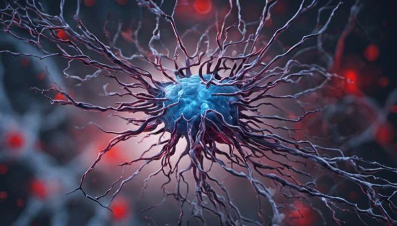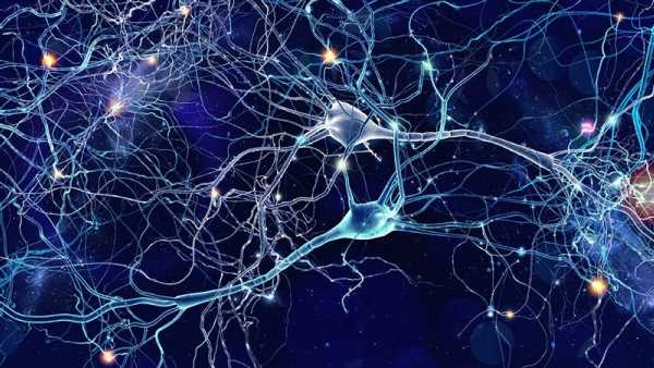
An often-overlooked communication strategy for neurons might be more prevalent than previously believed.
New research from biology professor Adam Miller’s lab in the UO’s College of Arts and Sciences illuminates the importance of neuron-to-neuron communication via direct electrical signaling, instead of the usual chemical messengers sent between cells. The team also identified proteins that might link disruptions in these electrical pathways to conditions like autism and epilepsy.
They describe their findings in a paper published May 11 in Current Biology.
Neurons are cells that send messages from the brain throughout the body. They direct everything an animal does: breathing, moving, thinking. The most well-known way that neurons signal is by releasing chemicals like dopamine and serotonin, which are then taken up by the next neuron in the communication chain. These connection points are called chemical synapses.
But Miller and his team are interested in a different kind of synapse: an electrical synapse. At an electrical synapse, neurons pass signals directly via electrical current, moving between cells through channels. Electrical synapses can form between many different parts of neurons and messages can flow through them in both directions, instead of just one way. Ultimately, neural circuits are created by the interactions between both electrical and chemical synapses.
Many neuroscientists previously thought that electrical synapses were most important during development but then were mostly phased out and replaced by chemical synapses, like crawling before learning to walk.
“But recent studies have found that electrical synapses persist throughout the brain, and they make up core parts of circuits themselves,” said Anne Martin, a postdoctoral researcher in Miller’s lab who led the new study.
Martin, Miller and their colleagues are trying to better understand how electrical synapses form and how they might affect the brain’s function.
In the latest paper, the team focused on the role of a protein called neurobeachin. They tested different versions of the protein in developing zebrafish and measured its effects on the electrical synapses.
Without neurobeachin working properly, the electrical synapses couldn’t form, the researchers found. Neurobeachin seems to function like a traffic controller, directing other proteins that are necessary for the synapse to work properly to the site of formation, Miller said. Without it, the proper components don’t end up in the right place, and the electrical messages can’t be sent.
Previous research has shown that neurobeachin also helps chemical synapses form. So the new research suggests a bridge between the two types of communication.
“People used to say those are distinct biochemical entities,” Miller said. “But now there’s this molecule that unites them in synapse formation.”
In future work, the team hopes to better understand how electrical and chemical synapses might relate to each other and the role they both play in neural circuits.
“We’re very interested in finding other bridges between electrical and chemical synapses,” Martin said. “We found one, neurobeachin, but we believe there could be others out there.”
They also plan to further explore the possible connections to human health. Miller’s team noticed behavioral changes in the fish with mutations in neurobeachin. And mutations in neurobeachin have previously been linked to autism and epilepsy, both conditions that involve changes in the way neurons talk to each other. Learning more about how neurobeachin affects neuron-to-neuron communication could help scientists better understand the origins of those brain differences.
More information:
E. Anne Martin et al, Neurobeachin controls the asymmetric subcellular distribution of electrical synapse proteins, Current Biology (2023). DOI: 10.1016/j.cub.2023.04.049
Journal information:
Current Biology
Source: Read Full Article
