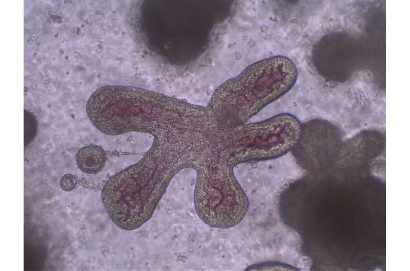
Experiments attempting to explain why bats can be infected with many viruses at a time without succumbing to diseases such as COVID-19—knowledge that could help us to reduce the threat to humans of infectious disease—have struggled until now with the fact that live wild bats make poor research subjects. To overcome this obstacle, for the first time researchers have grown rousette bat “organoids,” which reproduce intestines in vitro.
A paper describing the bat organoid growth technique appeared in the International Journal of Molecular Sciences on October 5.
Bats are the natural source of a raft of human pathogens (or, in epidemiological jargon, the “reservoir”—the host in which a pathogen survives without causing disease). These include the viruses that cause a great many illnesses such as Ebola, Marburg, Nipah, Hendra, SARS, MERS, and COVID-19. In fact, a single bat can host these viruses without getting sick. Why bats can live with so many viruses without themselves falling ill remains one of the great mysteries of virology and its neighboring disciplines. And solving this mystery has been made all the more urgent by the ongoing COVID-19 pandemic.
Yet bats are wild animals, not domesticated experimental animal subjects. It is much more difficult to conduct reproducible investigations on bats than on more common experimental animals such as mice or pigs. And so most experiments have had to take place on cell lines taken from bats rather than bats themselves or bat organs.
“If this experimental blockage could be overcome, virus-bat relationships could be understood and lead to reduce human illness and death.,” said Tsutomu Omatsu, one of the authors of the paper and an associate professor with the Center for Infectious Diseases of Epidemiology and Prevention Research at Tokyo University of Agriculture and Technology.
So the researchers developed a bat organoid that could be used for such experimentation. An organoid is a three-dimensional tissue construct grown “in vitro” (in a petri dish or with other laboratory equipment) from stem cells and that mimics the organ in the living animal. In this case, they grew organoids from cells from the intestine of a flying fox, the species Rousettus leschenaultia within the wider genus of Rousettus, also known as Rousette bats.
They chose Rousette bats, a type of megabat or fruit bat, because they are thought to be a natural reservoir of the filovirus family of viruses, including the Ebola and Marburg viruses. This particular species of rousette bat was also selected because in previous research, another species of rousette bat, Rousettus leschenaultii, had shown a transient but not robust infection from an experimental inoculation of SARS-CoV-2, the virus that causes COVID-19, while a cell line from the intestine of Rousettus leschenaultia had not been infected at all. Several species of flying foxes in Southeast Asia and Australia have also been found to be hosts of Pteropine orthoreovirus (PRV), which has caused respiratory disease in humans.
The researchers first had to find an optimum medium for the growth of bat intestine cells. They did this by attempting to culture organoids with nine different growth supplements (nutrients and other molecules that encourage cell proliferation). Three out of the nine achieved significantly higher cell growth and proliferation rates after seven days.
In addition, the rousette bat intestinal organoids grown with these three supplements were long-lived, being able to maintain active proliferation for up to ten passages (up to 10 times of reconstructions of organoids from separated cells that were composed of the previous organoids). Organoids that were long-term cryopreserved (in essence frozen) could also grow normally once thawed.
To confirm that the organoid was mimicking the epithelial (outer) tissue of the bat’s intestine—the part of the bat organ that first encounters virus particles and thus of particular scientific interest—the researchers deployed two techniques. First, they used transmission electron microscopy to investigate the cellular anatomy (histology) of the organoids. Second, they used immunofluorescence staining—a common method used to detect and visualize molecules in biological samples—to look for molecular markers that indicate that the tissue under investigation comes from bat intestines. Together, these two techniques told the researchers that the organoids were indeed recreating the typical cellular components of rousette bat intestine tissues.
An initial test of the use of the organoids to investigate viral relationships was also performed. They were shown to be susceptible to PRV but not to SARS-CoV-2 in experimental inoculation.
Source: Read Full Article
