Since coronavirus disease 2019 (COVID-19) first emerged, vast strides have been made in detection and testing technology. During the initial phases of the pandemic, while countries were forced to rely on ad-hoc PCR assays to confirm infection with the disease, most governments told anyone showing symptoms to isolate.
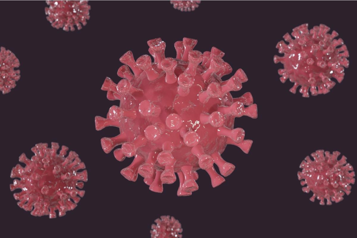
 Study: Nanobody-Functionalized Cellulose for Capturing SARS-CoV-2. Image Credit: Polina Tomtosova/Shutterstock
As the PCR assays became standardized and point-of-care lateral flow tests allowed rapid disease detection, testing was allowed before isolation. However, developing countries struggle with these expensive methods. In a study published in Applied and Environmental Microbiology, researchers from Northeastern University have developed a new, more cost-effective method for immobilizing severe acute respiratory coronavirus 2 (SARS-CoV-2).
The study
The researchers' core plan for creating a low-cost method for capturing and detecting SARS-CoV-2 required them to generate a bifunctional protein through the genetic fusion of high-affinity Nb—Ty1, which would target both the RBD of SARS-CoV-2 and the cellulose-binding domain (CBD).
To enable sampling and immobilization of SARS-CoV-2 from the environment or specimens from infected individuals, the scientists immobilized fusion proteins to the surface of filter papers and other cellulose materials. This should allow the bifunctional Nb-CBD to be immobilized in an orientation that should favor interactions between the Nb of interest and the antigen due to the specific interactions between the CBD domain and cellulose.
The bifunctional protein was integrated into a cellulose affinity purification column to allow for SARS-CoV-2 specific filtration in the hopes that this would prove that this strategy could reduce/eliminate viral load in blood products while maintaining blood viability.
The DNA for the Nb-CBD fusion protein was first cloned in E. coli, with the CBD placed at the C terminus of Nb to avoid steric hindrance. A his-tag was added to the N-terminus for metal affinity purification, and a FLAG epitope was inserted between Nb and the CBD. This epitope could act as both hydrophilic, flexible linkage and a tag for immunostaining.
To evaluate the effectivity of the fusion proteins for cellulose binding and Nb-specific target recognition, the scientists began by spotting and air drying purified fusion proteins on cellulose paper and then staining the paper with a rat antibody against the FLAG epitope. This was followed by a secondary ant-rat antibody conjugated with horseradish peroxide (HRP).
Incubation with 3'3-Diaminobenzidine (DAB) resulted in the visualization of a dark precipitate. Serially diluted fusion proteins were immobilized on the filter paper and immunostained with an anti-FLAG antibody to quantify the fusion protein's binding efficiency to the cellulose paper. They found that a surface area of 1mm2 could be saturated by 500ng of Nb-CBD proteins.
Speculating that the CBD's ability to act as a natural affinity ligand to cellulose could allow the E.coli cell lysate to be directly immobilized to filter paper and washed, avoiding the need for extensive and impractical protein purification later, the researchers began by incubating the cell lyase with filter discs. Non-specific proteins were removed via washing, and the functionalized filter discs were then subjected to media containing recombinant RBD. The filter paper captured the RBD, showing strong dark straining, whereas controls showed light or no staining.
To increase the surface density of the immobilized fusion protein, the researchers began by evaluating the capability of the fusion proteins to capture non-replicative lentivirus pseudotyped with the spike protein. They used both wild-type spike and spike protein carrying the D614G mutation, which confer worse symptoms and higher transmission.
They found that the Nb-CBD immobilized filter paper showed a 2-fold increase in the ability to capture the spike protein for both variants compared to filter paper alone and a 1.5 fold increase compared to Nb alone. To increase the capture efficiency, they attempted to incorporate the fusion proteins into regenerated amorphous cellulose, which has a higher surface area ratio than filter paper. Adding an additional washing step using Triton, the researchers also increased the capture efficiency for wild-type 3.5 fold and D614G variant by eightfold.
Conclusion
The authors highlight that they have created a simple, versatile, and affordable technology that effectively immobilizes SARS-CoV-2 on cellulose surfaces. This could be used as a wastewater surveillance sampling technology and a basic diagnostic platform. As scientists continue to warn that leaving large portions of the world unvaccinated and vulnerable to SARS-CoV-2 infection will continue to lead to the emergence of new variants, this could help developing countries effectively combat the disease.
-
Sun, X. et al., (2022). Nanobody-Functionalized Cellulose for Capturing SARS-CoV-2. Applied And Environmental Microbiology. doi: https://doi.org/10.1128/aem.02303-21 https://journals.asm.org/doi/10.1128/aem.02303-21
Posted in: Medical Science News | Medical Research News | Disease/Infection News
Tags: Antibody, Antigen, Blood, Cell, Coronavirus, Coronavirus Disease COVID-19, covid-19, Diagnostic, DNA, E. coli, Genetic, Lentivirus, Ligand, Lysate, Microbiology, Mutation, Pandemic, Protein, Protein Purification, Respiratory, SARS, SARS-CoV-2, Severe Acute Respiratory, Spike Protein

Written by
Sam Hancock
Sam completed his MSci in Genetics at the University of Nottingham in 2019, fuelled initially by an interest in genetic ageing. As part of his degree, he also investigated the role of rnh genes in originless replication in archaea.
Source: Read Full Article
